PHARYNX-I Presented by :- Dr. Sushma Tomar Associate

17 Slides2.29 MB
PHARYNX-I Presented by :- Dr. Sushma Tomar Associate Professor Department of Anatomy
Lesson Plan Introduction Boundaries Subdivisions Nasopharynx: Location. Communications. Pharyngeal Isthmus. Features. Applied Aspects.
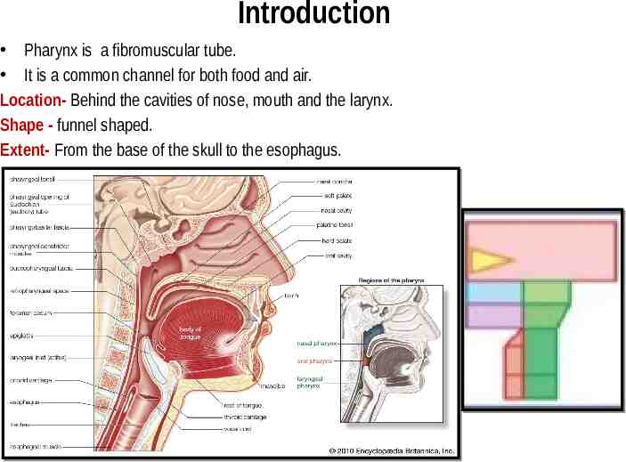
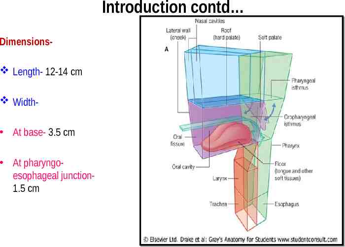
Introduction Pharynx is a fibromuscular tube. It is a common channel for both food and air. Location- Behind the cavities of nose, mouth and the larynx. Shape - funnel shaped. Extent- From the base of the skull to the esophagus.
Introduction contd Dimensions Length- 12-14 cm Width At base- 3.5 cm At pharyngoesophageal junction1.5 cm
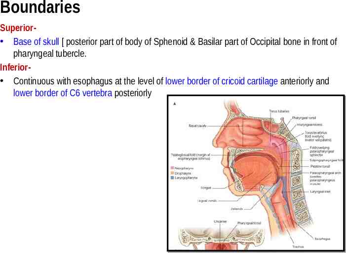
Boundaries Superior Base of skull [ posterior part of body of Sphenoid & Basilar part of Occipital bone in front of pharyngeal tubercle. Inferior Continuous with esophagus at the level of lower border of cricoid cartilage anteriorly and lower border of C6 vertebra posteriorly
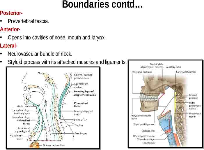
Boundaries contd Posterior Prevertebral fascia. Anterior Opens into cavities of nose, mouth and larynx. Lateral Neurovascular bundle of neck. Styloid process with its attached muscles and ligaments.
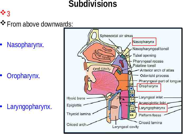
Subdivisions 3 From above downwards: Nasopharynx. Oropharynx. Laryngopharynx.
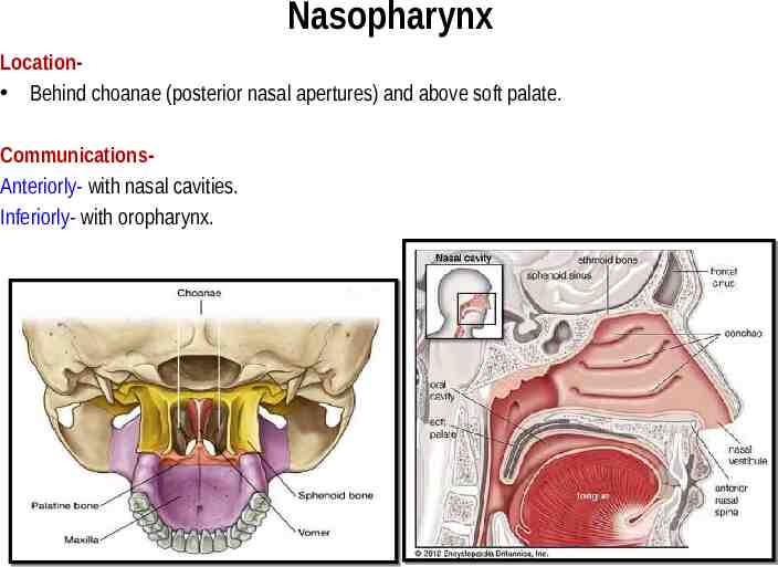
Nasopharynx Location Behind choanae (posterior nasal apertures) and above soft palate. CommunicationsAnteriorly- with nasal cavities. Inferiorly- with oropharynx.
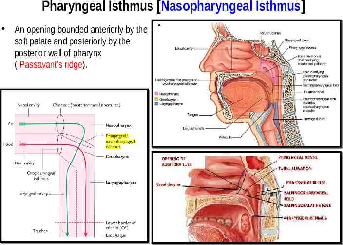
Pharyngeal Isthmus [Nasopharyngeal Isthmus] An opening bounded anteriorly by the soft palate and posteriorly by the posterior wall of pharynx ( Passavant’s ridge).
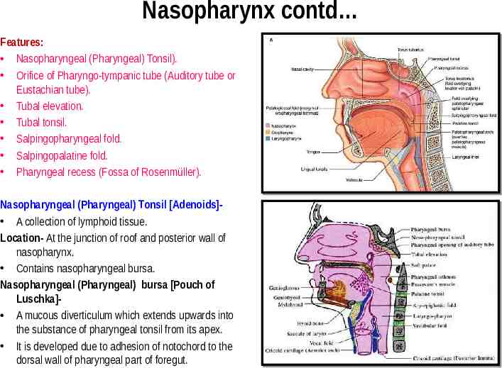
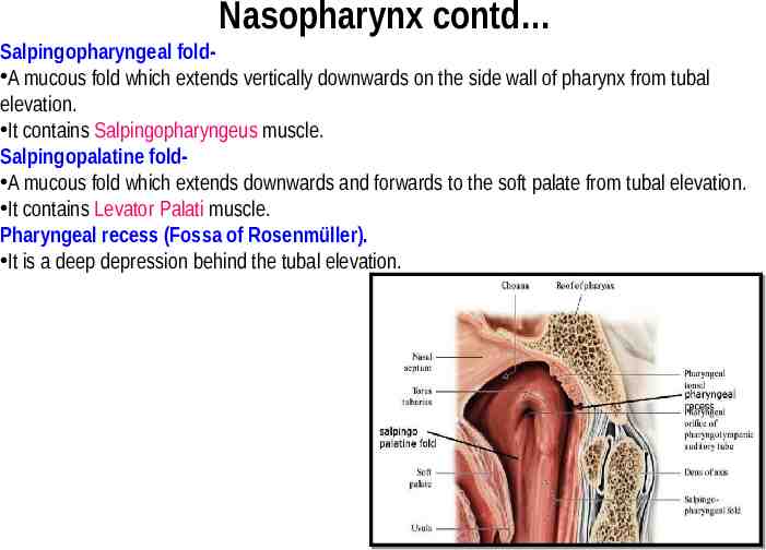
Nasopharynx contd Features: Nasopharyngeal (Pharyngeal) Tonsil). Orifice of Pharyngo-tympanic tube (Auditory tube or Eustachian tube). Tubal elevation. Tubal tonsil. Salpingopharyngeal fold. Salpingopalatine fold. Pharyngeal recess (Fossa of Rosenmüller). Nasopharyngeal (Pharyngeal) Tonsil [Adenoids] A collection of lymphoid tissue. Location- At the junction of roof and posterior wall of nasopharynx. Contains nasopharyngeal bursa. Nasopharyngeal (Pharyngeal) bursa [Pouch of Luschka] A mucous diverticulum which extends upwards into the substance of pharyngeal tonsil from its apex. It is developed due to adhesion of notochord to the dorsal wall of pharyngeal part of foregut.
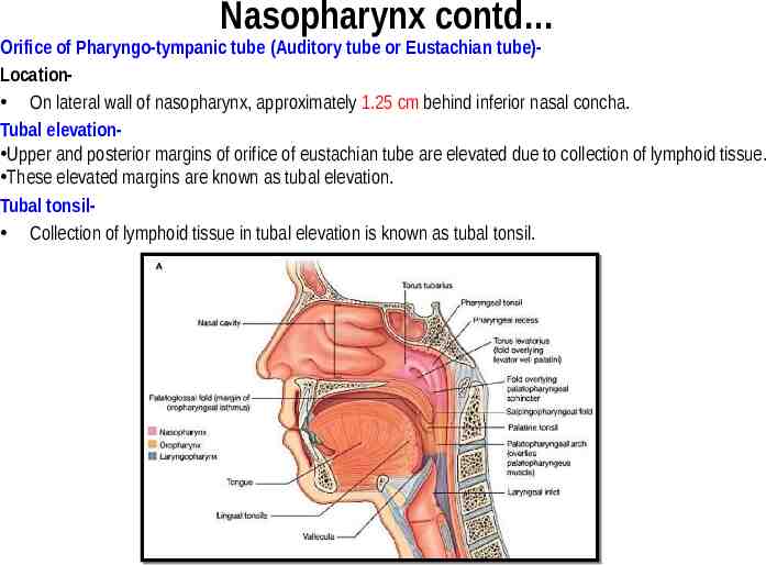
Nasopharynx contd Orifice of Pharyngo-tympanic tube (Auditory tube or Eustachian tube)Location On lateral wall of nasopharynx, approximately 1.25 cm behind inferior nasal concha. Tubal elevation Upper and posterior margins of orifice of eustachian tube are elevated due to collection of lymphoid tissue. These elevated margins are known as tubal elevation. Tubal tonsil Collection of lymphoid tissue in tubal elevation is known as tubal tonsil.
Nasopharynx contd Salpingopharyngeal fold A mucous fold which extends vertically downwards on the side wall of pharynx from tubal elevation. It contains Salpingopharyngeus muscle. Salpingopalatine fold A mucous fold which extends downwards and forwards to the soft palate from tubal elevation. It contains Levator Palati muscle. Pharyngeal recess (Fossa of Rosenmüller). It is a deep depression behind the tubal elevation.
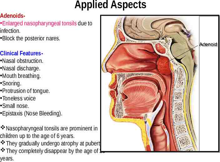
Applied Aspects Adenoids Enlarged nasopharyngeal tonsils due to infection. Block the posterior nares. Clinical Features Nasal obstruction. Nasal discharge. Mouth breathing. Snoring. Protrusion of tongue. Toneless voice Small nose. Epistaxis (Nose Bleeding). Nasopharyngeal tonsils are prominent in children up to the age of 6 years. They gradually undergo atrophy at puberty. They completely disappear by the age of 20 years.
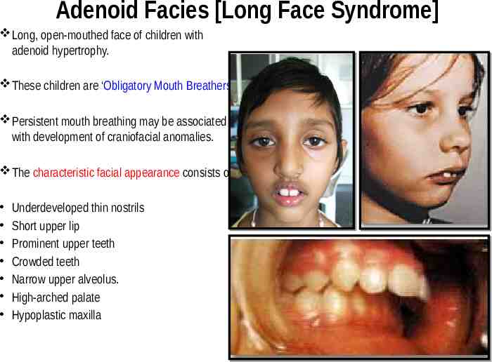
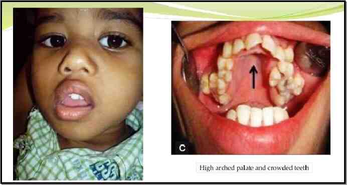
Adenoid Facies [Long Face Syndrome] Long, open-mouthed face of children with adenoid hypertrophy. These children are ‘Obligatory Mouth Breathers’. Persistent mouth breathing may be associated with development of craniofacial anomalies. The characteristic facial appearance consists of: Underdeveloped thin nostrils Short upper lip Prominent upper teeth Crowded teeth Narrow upper alveolus. High-arched palate Hypoplastic maxilla
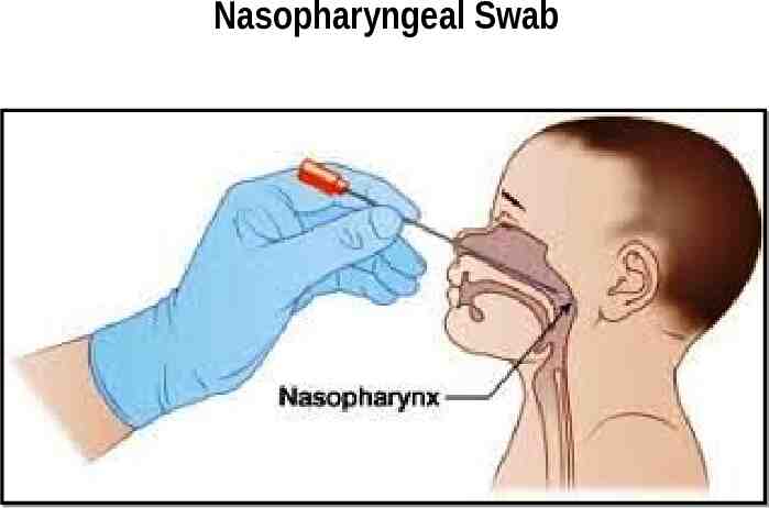
Nasopharyngeal Swab