Hypersensitivity Robert Beatty MCB150


48 Slides1.88 MB
Hypersensitivity Robert Beatty MCB150
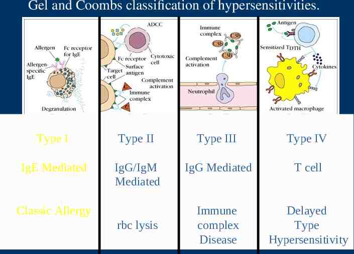
Gel and Coombs classification of hypersensitivities. Type I Type II Type III Type IV IgE Mediated IgG/IgM Mediated IgG Mediated T cell Immune complex Disease Delayed Type Hypersensitivity Classic Allergy rbc lysis
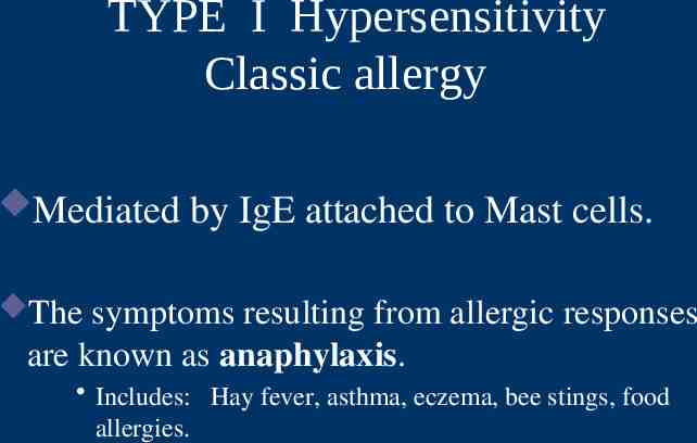
TYPE I Hypersensitivity Classic allergy Mediated by IgE attached to Mast cells. The symptoms resulting from allergic responses are known as anaphylaxis. Includes: Hay fever, asthma, eczema, bee stings, food allergies.
Allergens Allergens are nonparasite antigens that can stimulate a type I hypersensitivity response. Allergens bind to IgE and trigger degranulation of chemical mediators.
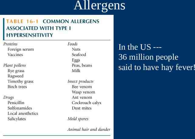
Allergens In the US --36 million people said to have hay fever!
Characteristics of allergens Small 15-40,000 MW proteins. Specific protein components – Often enzymes. Low dose of allergen Mucosal exposure. Most allergens promote a Th2 immune.
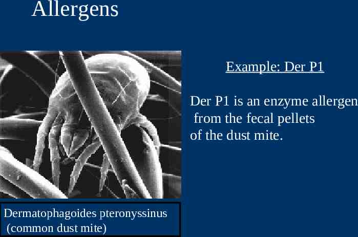
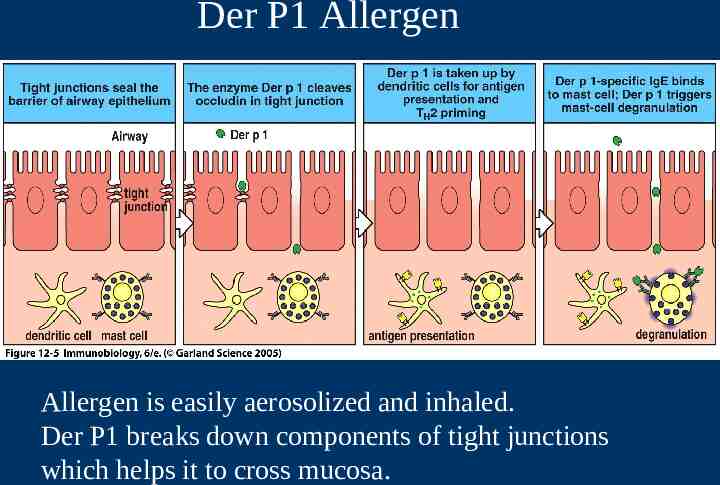
Allergens Example: Der P1 Der P1 is an enzyme allergen from the fecal pellets of the dust mite. Dermatophagoides pteronyssinus (common dust mite)
Der P1 Allergen Allergen is easily aerosolized and inhaled. Der P1 breaks down components of tight junctions which helps it to cross mucosa.
Atopy Atopy is the term for the genetic trait to have a predisposition for localized anaphylaxis. Atopic individuals have higher levels of IgE and eosinophils.
Genetic Predisposition Type I hypersensitivity Candidate polymorphic genes include: – IL-4 Receptor. – IL-4 cytokine (promoter region). – Fc RI. High affinity IgE receptor. – Class II MHC (present peptides promoting Th2 response). – Inflammation genes.
Mechanisms of allergic response Sensitization Repeated exposure to allergens initiates immune response that generates IgE isotype. Th2 cells required to provide the IL-4 required to get isotype switching to IgE.
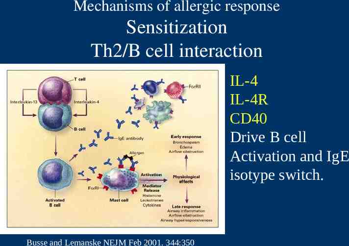
Mechanisms of allergic response Sensitization Th2/B cell interaction IL-4 IL-4R CD40 Drive B cell Activation and IgE isotype switch. Busse and Lemanske NEJM Feb 2001. 344:350
Mechanisms of allergic response Sensitization The IgE can attach to Mast cells by Fc receptor, which increases the life span of the IgE. Half-life of IgE in serum is days whereas attached to Fc R it is increased to months.
Mechanisms of allergic response Fc receptors (Fc R) Fc R1 high affinity IgE receptor found on – mast cells/basophils/activated eosinophils. Allergen binding to IgE attached to Fc R1 triggers release of granules from cell.
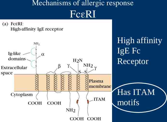
Mechanisms of allergic response Fc RI High affinity IgE Fc Receptor Has ITAM motifs
Mechanisms of allergic response Effector Stage of Hypersensitivity Secondary exposure to allergen Mast cells are primed with IgE on surface. Allergen binds IgE and cross-links to activate signal with tyrosine phosphorylation, Ca influx, degranulation and release of mediators.
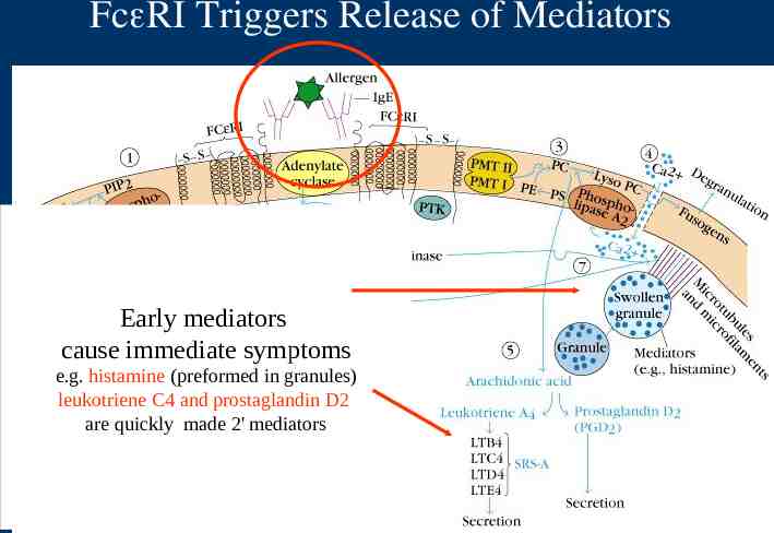
Fc RI Triggers Release of Mediators Early mediators cause immediate symptoms e.g. histamine (preformed in granules) leukotriene C4 and prostaglandin D2 are quickly made 2' mediators
Mediators of Type I Hypersensitivity Immediate effects Histamine – Constriction of smooth muscles. Bronchiole constriction wheezing. Constriction of intestine cramps-diarrhea. – Vasodilation with increased fluid into tissues causing increased swelling or fluid in mucosa. – Activates enzymes for tissue breakdown. Leukotrienes Prostaglandins
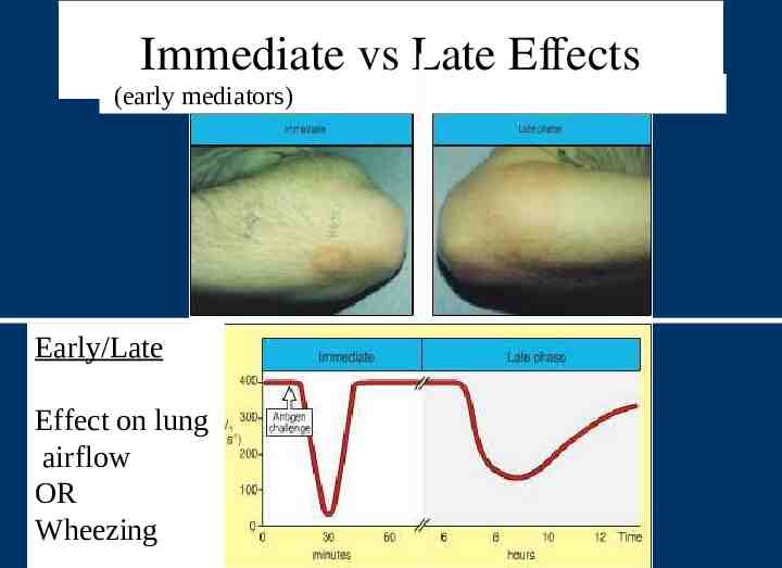
Immediate vs Late Effects (early mediators) Early/Late Effect on lung airflow OR Wheezing
Mediators of Type I Hypersensitivity Primary Mediators Pre-formed mediators in granules Histamine Cytokines TNF- , IL-1, IL-6. Chemoattractants for Neutrophils and Eosinophils. Enzymes – tryptase, chymase, cathepsin. – Changes in connective tissue matrix, tissue breakdown.
Type I Hypersensitivity Secondary mediators Mediators formed after activation Leukotrienes Prostaglandins Th2 cytokines- IL-4, IL-5, IL-13, GM-CSF
Continuation of sensitization cycle Mast cells control the immediate response. Eosinophils and neutrophils drive late or chronic response. More IgE production further driven by activated Mast cells, basophils, eosinophils.
Continuation of sensitization cycle Eosinophils Eosinophils play key role in late phase reaction. Eosinophils make – enzymes, – cytokines (IL-3, IL-5, GM-CSF), – Lipid mediators (LTC4, LTD4, PAF) Eosinophils can provide CD40L and IL-4 for B cell activation.
Localized anaphylaxis Target organ responds to direct contact with allergen. Digestive tract contact results in vomiting, cramping, diarrhea. Skin sensitivity usually reddened inflamed area resulting in itching. Airway sensitivity results in sneezing and rhinitis OR wheezing and asthma.
Systemic anaphylaxis Systemic vasodilation and smooth muscle contraction leading to severe bronchiole constriction, edema, and shock. Similar to systemic inflammation.
Treatment for Type I Pharmacotherapy Drugs. – Non-steroidal anti-inflammatories – Antihistamines block histamine receptors. – Steroids – Theophylline OR epinephrine -prolongs or increases cAMP levels in mast cells which inhibits degranulation.
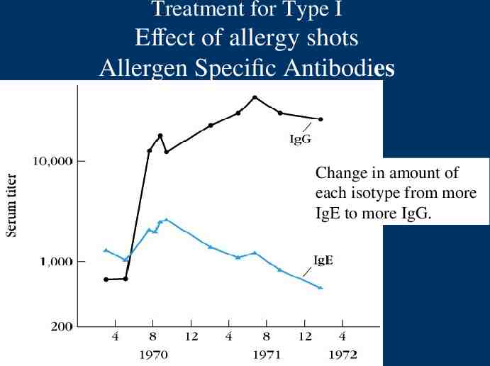
Treatment for Type I Immunotherapy – Desensitization (hyposensitization) also known as allergy shots. – Repeated injections of allergen to reduce the IgE on Mast cells and produce IgG.
Treatment for Type I Effect of allergy shots Allergen Specific Antibodies Change in amount of each isotype from more IgE to more IgG.
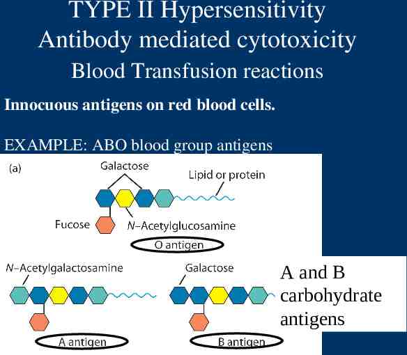
TYPE II Hypersensitivity Antibody mediated cytotoxicity Blood Transfusion reactions Innocuous antigens on red blood cells. EXAMPLE: ABO blood group antigens A and B carbohydrate antigens
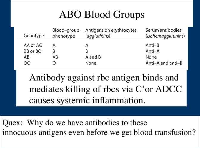
ABO Blood Groups Antibody against rbc antigen binds and mediates killing of rbcs via C’or ADCC causes systemic inflammation. Quex: Why do we have antibodies to these innocuous antigens even before we get blood transfusion?
TYPE II Antibody mediated cytotoxicity Drug reactions Drug binds to rbc surface and antibody against drug binds and causes lysis of rbcs. Immune system sees antibody bound to "foreign antigen" on cell. ADCC
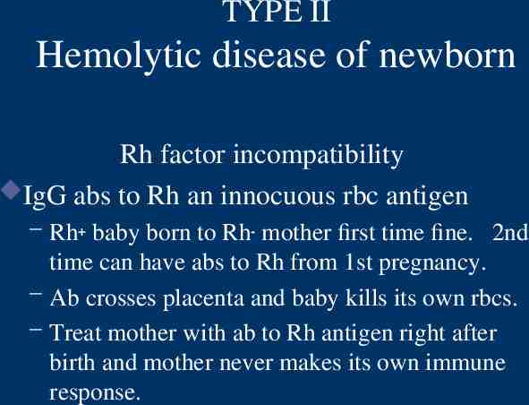
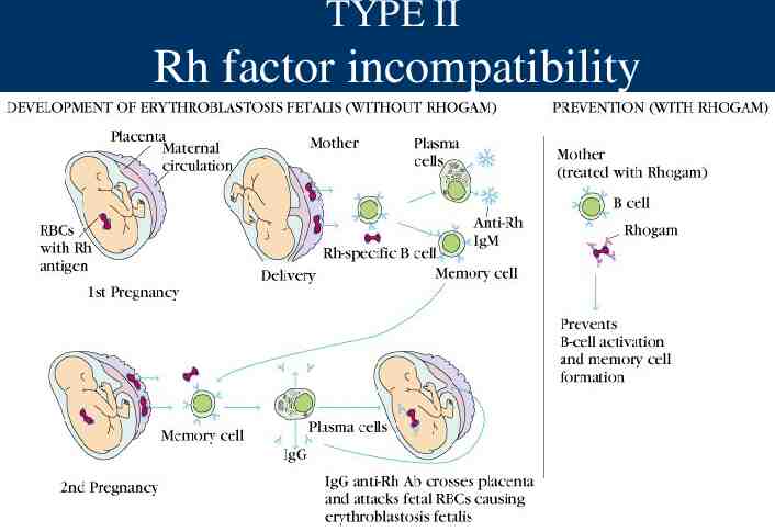
TYPE II Hemolytic disease of newborn Rh factor incompatibility IgG abs to Rh an innocuous rbc antigen – Rh baby born to Rh- mother first time fine. 2nd time can have abs to Rh from 1st pregnancy. – Ab crosses placenta and baby kills its own rbcs. – Treat mother with ab to Rh antigen right after birth and mother never makes its own immune response.
TYPE II Rh factor incompatibility
TYPE III Antigen antibody immune complexes. IgG mediated Immune Complex Disease Large amount of antigen and antibodies form complexes in blood. If not eliminated can deposit in capillaries or joints and trigger inflammation.
TYPE III Immune Complexes PMNs and macrophages bind to immune complexes via FcR and phagocytize the complexes. BUT If unable to phagocytize the immune complexes can cause inflammation via C’ activation --- C3a C4a, C5a and "frustrated phagocytes".
TYPE III Immune Complex Disease "Frustrated Phagocytes" If neutrophils and macrophages are unable to phagocytize the immune complexes these cells will degranulate in the area of immune complex deposition and trigger inflammation. Unable to eat -------try to digest outside cell.
TYPE III Immune Complex Disease Localized disease Deposited in joints causing local inflammation arthritis. Deposited in kidneys glomerulonephritis.
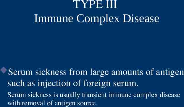
TYPE III Immune Complex Disease Serum sickness from large amounts of antigen such as injection of foreign serum. Serum sickness is usually transient immune complex disease with removal of antigen source.
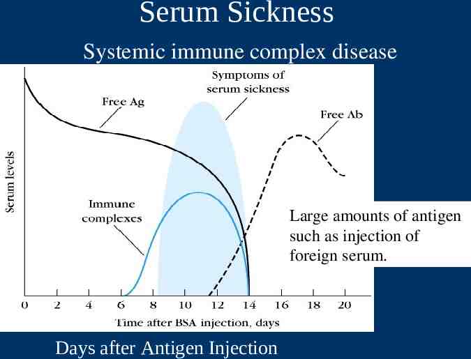
Serum Sickness Systemic immune complex disease Large amounts of antigen such as injection of foreign serum. Days after Antigen Injection
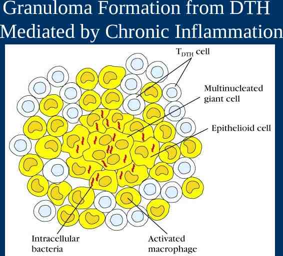
Delayed type hypersensitivity Th1 cells and macrophages DTH response is from: – Th1 cells release cytokines to activate macrophages causing inflammation and tissue damage. – Continued macrophage activation can cause chronic inflammation resulting in tissue lesions, scarring, and granuloma formation. Delayed is relative because DTH response arise 24-72 hours after exposure rather than within minutes.
Stages of Type IV DTH Sensitization stage Memory Th1 cells against DTH antigens are generated by dendritic cells during the sensitization stage. These Th1 cells can activate macrophages and trigger inflammatory response.
Stages of Type IV DTH Effector stage Secondary contact yields what we call DTH. Th1 memory cells are activated and produce cytokines. – IFN- , TNF- and TNF- which cause tissue destruction, inflammation. – IL-2 that activates T cells and CTLs. – Chemokines- for macrophage recruitment. – IL-3, GM-CSF for increased monocyte/macrophage
Stages of Type IV DTH Effector stage Secondary exposure to antigen Inflamed area becomes red and fluid filled can form lesion. – From tissue damage there is activation of clotting cascades and tissue repair. Continued exposure to antigen can cause chronic inflammation and result in granuloma formation.
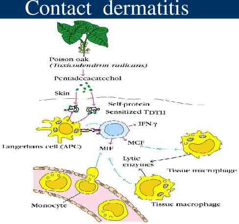
Type IV DTH Contact dermatitis The response to poison oak is a classic Type IV. – Small molecules act as haptens and complex with skin proteins to be taken up by APCs and presented to Th1 cells to get sensitization. – During secondary exposure Th1 memory cells become activated to cause DTH.
Contact dermatitis
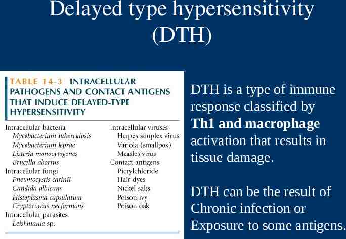
Delayed type hypersensitivity (DTH) DTH is a type of immune response classified by Th1 and macrophage activation that results in tissue damage. DTH can be the result of Chronic infection or Exposure to some antigens.
Granuloma Formation from DTH Mediated by Chronic Inflammation
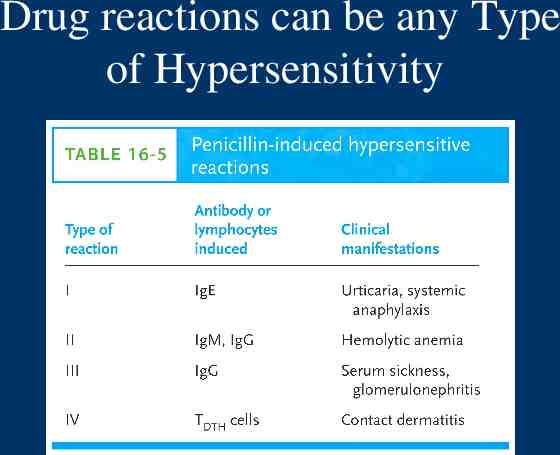
Drug reactions can be any Type of Hypersensitivity