Chapter 3: Cells – The Living Units


75 Slides2.37 MB
Chapter 3: Cells – The Living Units
Cellular Basis of Life Robert Hooke – late 1600s – observed plant cells 1830s Schleiden & Schwann – concluded all living things are composed of cells Rudolf Virchow – found – all cells come from preexisting cells
1800s- Cell Theory 1. Cell is the basic structural and functional unit of living organisms 2. Activity of organism – depends on individual and collective activities of cells 3. Principal of complementary structure and function – biochemical activities of cells are dictated by relative numbers of subcellular structures 4. Continuity of life from one generation to another has a cellular basis
Human Body Trillions of cells 200 different cell types Size ranges from 2 µm to 1 m Structural unit of all living things All cells composed of C, N, O, and other trace elements All cells have same basic parts (generalized or composite cell)
Human Cells All have 3 main parts 1. Plasma membrane – outer boundary 2. Cytoplasm – intracellular fluid packed with organelles 3. Nucleus – controls cellular activates
Plasma Membrane Also called the cell membrane Flexible Defines the extent of cell Separates intracellular fluid from extracellular fluid
Fluid Mosaic Model Thin (7-10 nm) structure Composed of double layer – bilayer of lipid molecules with protein molecules dispersed in it
Membrane Lipids Phospholipids – Polar head – hydrophilic – “water loving” Non-polar tail – hydrophobic – “water fearing” Polar heads are attracted to water, lie on inner and out surfaces Sandwich like structure – heads on outside, tails on inside Majority of tails – unsaturated – have kinks and increase membrane fluidity
Membrane Lipids Glycoproteins - lipids with sugars attached – Only on outer membrane surface – 5 % of total membrane Cholesterol – 20 % of total membrane – Polar region hydroxyl group – Nonpolar region – fused ring – Stabilizes membrane – Increases mobility of phospholipids Lipid Rafts – 20 % of outer membrane – Assemblies of saturated phospholipids – Packed tightly together – Platforms for receptor molecules and cell signaling
Membrane Proteins Make up approximately 50 % of membrane Responsible for specialized function 2 kinds of proteins – Integral proteins – Peripheral proteins
Membrane Proteins – Integral Protiens Inserted into membrane Some stick out only on one side Others span entire membrane – transmembrane proteins – Hydrophobic and hydrophilic regions – Most involved in transport – Form pores or channels Others – carriers – bind substances and move them into membrane Also receptors – relay messages to interior of cell
Membrane Proteins – Peripheral Proteins Not embedded in membrane Attached loosely to integral proteins Include filaments that support membrane Some are enzymes Others – motor proteins – change cell shape during division and cell contraction Glycocaylx – “sugar coating” sugars attached to proteins Highly specific biological markers – allow cells to recognize each other
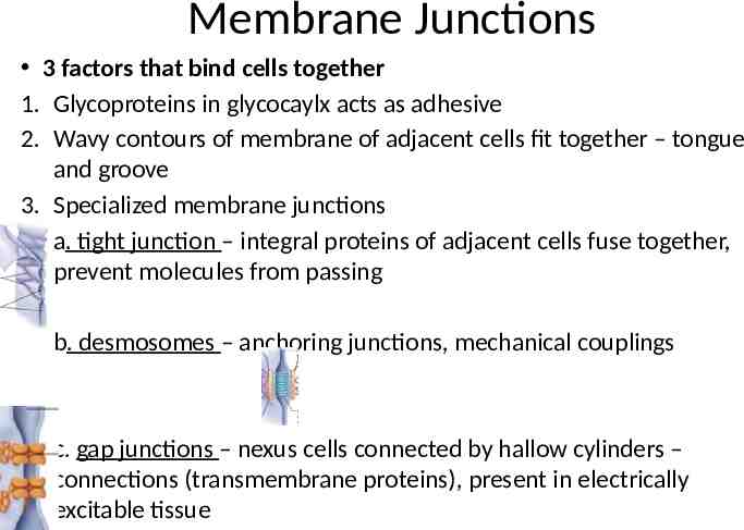
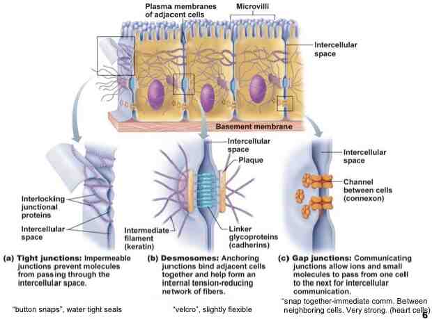
Membrane Junctions 3 factors that bind cells together 1. Glycoproteins in glycocaylx acts as adhesive 2. Wavy contours of membrane of adjacent cells fit together – tongue and groove 3. Specialized membrane junctions a. tight junction – integral proteins of adjacent cells fuse together, prevent molecules from passing b. desmosomes – anchoring junctions, mechanical couplings c. gap junctions – nexus cells connected by hallow cylinders – connections (transmembrane proteins), present in electrically excitable tissue
Membrane Transport Extracellular fluid – interstitial fluid Rich, nutritious “soup” Amino acids, sugars, fatty acids, vitamins, hormones, salts, wastes, etc. Plasma Membrane – selectively permeable Allows some substances to pass, but NOT others Passive Processes – substances cross without energy input Active Process – substances need energy input to cross
Transport - Passive Diffusion – movement of molecules from an area of high concentration to an area of low concentration Down the concentration gradient Kinetic energy of molecules moves them Speed of diffusion – influenced by: size – smaller the molecules are the faster they move Molecules move until equilibrium is reached (no net movement) Plasma membrane – physical barrier – but molecule will diffuse if it is– 1. Lipid soluble 2. Small enough to pass through channels 3. Assisted by carrier
Transport - Passive Simple Diffusion – nonpolar and lipid soluble substances diffuse directly though membrane Oxygen, carbon dioxide, fat-soluble vitamins, etc,
Transport - Passive Facilitated Diffusion – molecules that can not pass through the membrane by themselves Transported with the help of a protein Substance can binds the carrier protein in membrane Substance may also move through water filled channels Carriers – integral proteins – allow substances to pass through membrane Channels – transport proteins Transport water or ions through aqueous channels
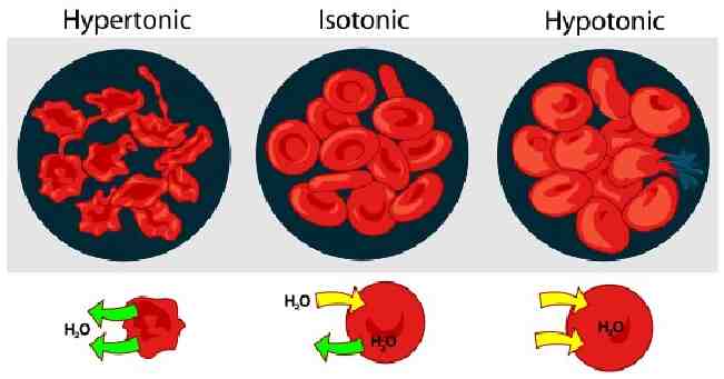
Transport - Passive Osmosis – diffusion of solvent (water) through a selectively permeable membrane Aquaporins (APQs) water specific channels in the membrane Moves down the concentration gradient Also depends on the concentration of solutes Osmolarity – total concentration of all solute particles in a solution
Transport Passive Osmosis cont – Water diffuses until hydrostatic pressure (back pressure exerted by water against the membrane) with in the cell is equal to its osmotic pressure (tendency of water to move into cell by osmosis) Tonicity – change in shape or tone of cells by altering internal water volume
Solutions Isotonic – cells with the same concentration of solutes on the inside and outside Hypertonic – solution has a higher concentration than the inside of the cell – Water moves out, cells shrinks Hypotonic – solution is more dilute than inside of cell – Water moves in, cell swells
Active Transport Processes Active Transport – requires protein Combines specifically and reversibly with transported substances Solute pumps – move solutes against the concentration gradient Requires the input of energy Symbort System – 2 substances transported the same way (both inside or both outside) Antiport System – 2 substances transported opposite ways (one inside and the other outside)
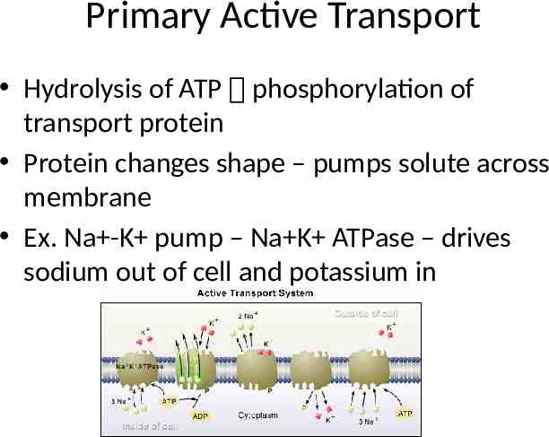
Primary Active Transport Hydrolysis of ATP phosphorylation of transport protein Protein changes shape – pumps solute across membrane Ex. Na -K pump – Na K ATPase – drives sodium out of cell and potassium in
Secondary Active Transport Single ATP powered pump indirectly drives secondary active transport Na moves back into cell, another substance is cotransported with it Ex. Sugar, amino acids, ions, etc
Vesicular Transport Fluids containing large particles and macromolecules are transported in membranous sacs – vesicles Exocytosis – process that ejects substances from interior of cell
Vesicular Transport Endocytosis – process that moves substances into the cell Substances moved in by the infolding of the membrane – coated pit – clathrin – protein coating, then vesicle detaches Phagocytosis – cells engulfs large solid material – Bacteria, debris, etc – Endocytic vesicle – phagosome – Amoeboid motion – flowing of cytoplasm into temporary pseudopods Pinocytosis – fluid phase endocytosis – Plasma membrane surrounds small volume of fluid containing dissolved material Receptor mediated endocytosis – plasma membrane binds only certain substances
Vesicular Transport Other protein coats – Caleolae – tubular or flask shaped inpocketings of plasma membrane Coatomer (COP1 & COP2) proteins – vesicular trafficking Transport substances between organelles
Plasma Membrane – Membrane Potential Membrane Potential – voltage – electrical potential energy resulting from separation of oppositely charged particles
Plasma Membrane – Membrane Potential Resting State – resting membrane potential – range -50 -100mV Cell said to be polarized (-) indicates inside is negative compared to the outside Diffusion – causes ionic imbalances that polarize the membrane, active transport maintains membrane polarization Ions – K and protein – inside cell Na and Cl- outside cell Membrane somewhat permeable to K - leaky channels Protein anions cannot follow
Plasma Membrane – Membrane Potential Membrane becomes negative -90 mV Na also a factor – attracted to cell interior – bring membrane to -70 mV Active transport – depends on diffusion More Na in, the more is pumped out Na /K pump – 3 Na out for 2 K in Electrochemical gradient – electrical and concentration (chemical) forces
Cell-Environment Interactions Cell Adhesion Molecules (CAMs) Key role in embryonic development, wound repair, and immunity Sticky glycoproteins
Cell-Environment Interactions Functions – 1. Molecular “Velcro” cells use to anchor themselves to molecules in extracellular space and to each other 2. The “arms: that migrating cells use to haul themselves past one another 3. SOS signals – sticking out from blood vessels lining that rally WBC to infected or damaged area 4. Mechanical sensors – respond to tension at cell surface by stimulating synthesis or degradation of adhesive membrane junction 5. Transmitters of intracellular signals that direct cell migration, proliferation, and specialization
Roles Membrane receptors – integral proteins and glycoproteins that serve as binding sites 1. Cell Signaling – coming together and touching of cells - Cells recognize each other - Essential for normal development and immunity
Roles 2. Chemical Signaling – - Ligands – signaling chemicals, bind to specific plasma membrane receptors - Include – neurotransmitters, hormones, paracrines - G-Protein – linked receptors – exert effects through G-protein - Second messengers are generated and connect plasma membrane events to internal - Ex. Cyclic AMP – Ca2 - Activates a protein kinase cascade - Nitric oxide (NO) – another messenger – important signaling molecule
Cytoplasm “cell forming’ material Cell material between the plasma membrane and the nucleus 3 major elements – cytosol, organelles, and inclusions
Cytoplasm 1. Cytosol Viscous semitransparent fluid Colloid and solution properties Dissolved (in water) – protein, sugar, salts, and solutes 2. Organelles Carry out specific functions – synthesize proteins, package proteins, etc. Chemical substances not always present 3. Inclusions Stored nutrients, lipid droplets, pigment, water containing vacuoles, crystals of various types
Organelles “little organs” Nonmembranous – lack membranes Membranous – with membranes
Mitochondria Lozenge-shaped membranous organelles Power plants of cells 2 membranes – – Outer – smooth and featureless – Inner – folds inward forming cristae Gel-like substance inside Food particles – glucose – broken down into water and carbon dioxide by enzymes Metabolites broken down and oxidized – energy released and captured Aerobic cellular respiration
Mitochondria Contain their own DNA, RNA, and ribosomes Can reproduce themselves 37 genes – direct synthesis of 1% of proteins needed Similar to bacteria (purple bacteria phylum) Believed to have arose from bacteria that evaded plant and animal cells
Ribosomes Small dark staining granules Protein and a variety of RNA – ribosomal RNA Site of protein synthesis Some float freely Others attached to rough ER Free ribosomes – make soluble proteins Rough ER – make proteins destined for cell membranes or export
Endoplasmic Reticulum (ER) “network” with in cytoplasm Intracellular connected tubes and parallel membranes enclosed fluid filled cavities or cisternae Continuous with the nuclear membrane Half of cells total membrane 2 types – smooth and rough
Rough Endoplasmic Reticulum (RER) Surface covered with ribosomes Proteins from ribosomes – enter and are modified “Membrane factory”
Smooth Endoplasmic Reticulum (SER) Continuous with the rough ER No role in protein synthesis Involved in: 1. Lipid metabolism, cholesterol synthesis, synthesis of lipid components – of lipoproteins 2. Synthesis of steroid hormones (sex hormones) 3. Absorption, synthesis, and transport of fats 4. Detox of drugs, pesticides, and carcinogens 5. Breakdown of stored glycogen into free glucose - Muscle – also stores calcium
Golgi Apparatus Stacked and flattened membranous sacs Traffic director for cellular proteins Modify, concentrate and package proteins and lipids Secretory vesicles or granules migrated to plasma membrane and discharge contents
Lysosomes Inactive digestive enzymes Cells demolition crew 1. Digest particles taken in by endocytosis, particularly bacteria, viruses, toxins 2. Degrade worn-out or nonfunctional organelles 3. Metabolic functions – glycogen breakdown and release 4. Breakdown of nonuseful tissues – uterine lining during menstruation 5. Breaking down of bone to release calcium into blood - Autolysis – lysosome rupture and cell digests itself
Endomembrane System System of organelles that work together to: 1. Produce, store, and export biological materials 2. Degrade potential harmful substances ER, Golgi, secretory vessels, and lysozomes, also nuclear membrane
Peroxisomes “peroxide bodies” Membranous sacs with a variety of powerful enzymes Oxidases and catalyzes Oxidase – use molecular oxygen to detoxify harmful substances – alcohol/fermaldehyde Neutralize dangerous free radicals – highly reactive chemicals with unpaired electrons Convert to hydrogen peroxide Numerous in liver and kidney
Cytoskeleton “cell skeleton” Elaborate network of rods running through cytosol 3 parts – microfilaments, intermediate filaments, and microtubules
Cytoskeleton - Microtubules Elements with largest diameter Hallow tubes made of spherical protein subunits- tubulins Organelles attached Motor proteins – move and reposition organelles
Cytoskeleton - Microfilaments Thinnest elements of cytoskeleton Strands of protein – actin Act together with myosin to generate contractile forces
Cytoskeleton – Intermediate Filaments Though insoluble protein fibers Woven ropes Internal wires to resist pulling forces acting on cell
Centrosome and Centrioles Centrosome – cell center, microtubule organizing center Centrioles – small barrel shaped organelles oriented at right angles to each other
Cellular Extensions Cilia – whip like motile cellular extensions on cell surface Move substances in one direction across cell surface Flagella – long projection Ex. Sperm cell – commonly only a tail Bases – basal bodies – 9 2 pattern of microtubules Micorvilli – minute, fingerlike extensions of plasma membrane, increase the SA of plasma membrane
Nucleus Control center Genes All body cells have nucleus RBC eject their nucleus – anucleate Average – 5 µm Largest organelle 3 regions – nuclear envelope, nucleoli, and chromatin
Nuclear Envelope Double membrane bilayer Outer membrane continuous with ER Inner membrane lined by nuclear lamina Nuclear pores – protein (NPC) – aqueous transport channel – entry and exist Selectively permeable membrane Nucleoplasm – jellylike fluid on inside
Nucleoli Dark-staining spherical bodies with in the nucleus Typically 1 or 2 but maybe more Sites where ribosomal subunits are assembled Associated with the nucleolar organizer regions – genetic information for synthesizing rRNA rRNA – combined with proton ribosome
Chromatin Fine, unevenly stained network (microscope) Really bumpy threads Composed of 30: DNA, 60% histone proteins, 10% RNA chains Fundamental unit – nucleosomes – cluster of 8 histone proteins connected by DNA Pack DNA Ready to divide – condense into chromosomes
Cell Life Cycle Series of changes a cell goes through 2 major periods 1. Interphase 2. Cell Division
Interphase Period from cell formation to cell division Subphases: G1 (gap 1) phase – – Cell metabolically active – Synthesizes protein and grows S phase – DNA replicated – New histones made and assembled into Chromatin G2 (gap 2) phase – brief – Enzymes and proteins needed for division are synthesized
Interphase – DNA Replication 1. DNA helix unwinds 2. Helicase enzyme untwists helix, replication fork – Y-shaped separation 3. Each strand serves as a template 4. DNA Polymerase – attaches complementary nucleotides, leading strand and lagging strand 5. DNA ligase hooks segments of lagging strand together, leading strand - continuous
Cell Division M (mitotic) phase 2 events – mitosis and cytokinesis Mitosis – division of the nucleus 4 phases 1. 2. 3. 4. Prophase Metaphase Anaphase Telophase
Cell Division Cytokinesis – division of the cytoplasm Begins in late anaphase Plasma membrane drawn inward (cleavage furrow) by contractile ring 2 cells pinched apart
Control of Cell Cycle Signals for cell division 1. Ratio of SA to volume - 64 fold increase in volume, only 16 fold increase in SA - Inadequate SA for nutrient and waste exchange 2. Chemical Signals – growth factors, hormones, etc.
Control of Cell Cycle Cyclins and Cdks (cyclin dependent kinases) Proteins and enzymes that signal the cell to divide Also checkpoints for cell division – MPF – Mphase promoting factor – OK – signal to pass G2 and enter M
Protein Synthesis DNA – blueprint for proteins Gene - sequence of DNA that carries instructions for proteins DNA bases – A, C, T, G 3 bas segments – triplet – “word” that specifies an amino acid Most genes have exons – which code for amino acids separated by introns – noncoding segments of DNA Introns – can range from 60 100,000 nucleotides long – “junk DNA” – must be cut out to make a protein
Protein Synthesis DNA can not leave the nucleus so need a carrier RNA – (carrier) 3 types 1. Messenger RNA (mRNA) – long nucleotide strand, “half DNA” codes for a protein 2. Ribosomal RNA (rRNA) – part of ribosome 3. Transfer RNA (tRNA) – small roughly L-shaped molecules
Protein Synthesis 2 Parts 1. Transcription – DNA mRNA - initiation, elongation and termination - Promoter – start point - RNA polymerase – initiates transcription, pulls strands apart, aligns RNA nucleotides - Termination signal – transcription ends – mRNA pulls off
Protein Synthesis mRNA – must then be modified – splicosomes – cut introns (junk) and splice (glue together) remaining exons
Protein Synthesis 2. Translation – - base sequence (mRNA) converted to amino acids (protein) - Codon – 3 bas sequence on mRNA – 64 codons for 20 amino acids - Anticodon - 3 base sequence on tRNA – brings in amino acid - Amino acids hook together – form a chain PROTEIN
DNA – other roles Antisense RNA – can intercept and bind to protein coding mRNA, prevent it from being translated into protein microRNAs – small RNAs that can interfere and suppress mRNA Ribositches – folded mRNA, code for a protein – Can turn on protein synthesis in response to changes in the environment
Protein Degradation Proteins - no longer useful Ubiquitins – proteins that attach to old/bad protein – mark it for destruction Proteasomes – digest the protein
OUTSIDE Extracellular Materials Body fluids – interstitial fluid, blood plasma, cerebrospinal fluid Cellular secretions – substances that aid in digestion and act as lubricants Extracellular Matrix – jelly-like, composed of proteins and polysaccharides “cell glue” holds cells together
Development Embryo –cells – chemical signals that direct pathways of development Cell differentiation – development of specific and distinctive features of cells Apoptosis -programmed cell death Hyperplasia – accelerated growth Atrophy – decrease in size of organ or body tissue
Development Aging – Telomere – string of nucleotides at the end of a chromosome that protect it from fraying Get shorter with each cell division Aging – shorten, not protected Telomerase – enzyme that protects from degradation Found in egg and sperm cells, NOT in normal body cells “fountain of youth”