BOT3015L Symbioses Presentation created by Danielle Sherdan All
24 Slides6.52 MB
BOT3015L Symbioses Presentation created by Danielle Sherdan All photos from Raven et al. Biology of Plants except when otherwise noted
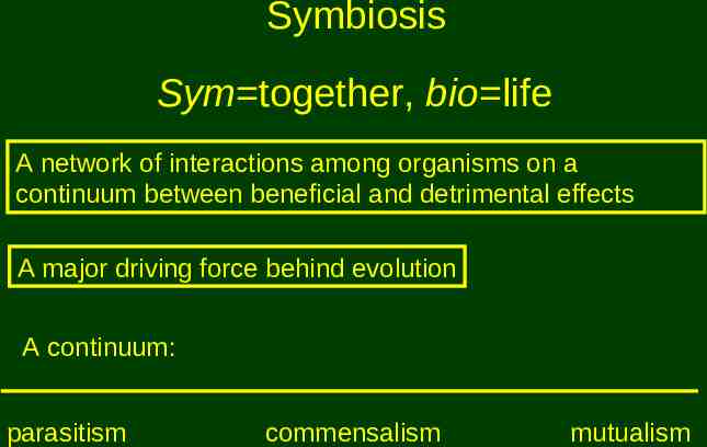
Symbiosis Sym together, bio life A network of interactions among organisms on a continuum between beneficial and detrimental effects A major driving force behind evolution A continuum: parasitism commensalism mutualism
A few examples Flowering plants and pollinating animals Humans and domesticated plants and animals Humans and bacteria in their digestive system Endosymbionts Origination of mitochondria and chloroplasts
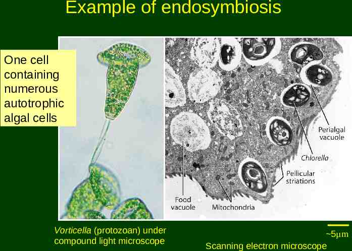
Example of endosymbiosis One cell containing numerous autotrophic algal cells Vorticella (protozoan) under compound light microscope 5µm Scanning electron microscope
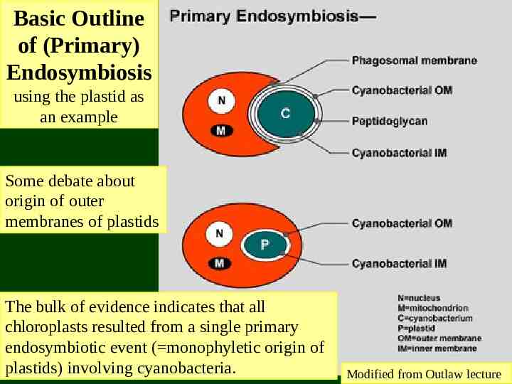
Basic Outline of (Primary) Endosymbiosis using the plastid as an example Some debate about origin of outer membranes of plastids The bulk of evidence indicates that all chloroplasts resulted from a single primary endosymbiotic event ( monophyletic origin of plastids) involving cyanobacteria. Modified from Outlaw lecture
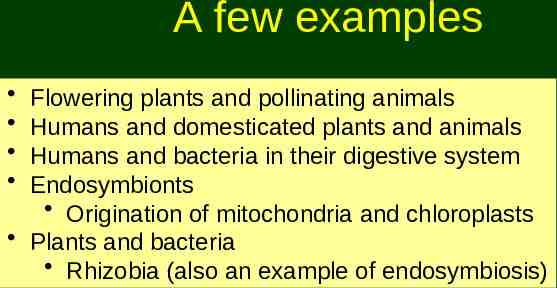
A few examples Flowering plants and pollinating animals Humans and domesticated plants and animals Humans and bacteria in their digestive system Endosymbionts Origination of mitochondria and chloroplasts Plants and bacteria Rhizobia (also an example of endosymbiosis)
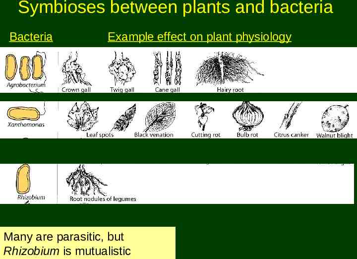
Symbioses between plants and bacteria Bacteria Example effect on plant physiology Many are parasitic, but Rhizobium is mutualistic
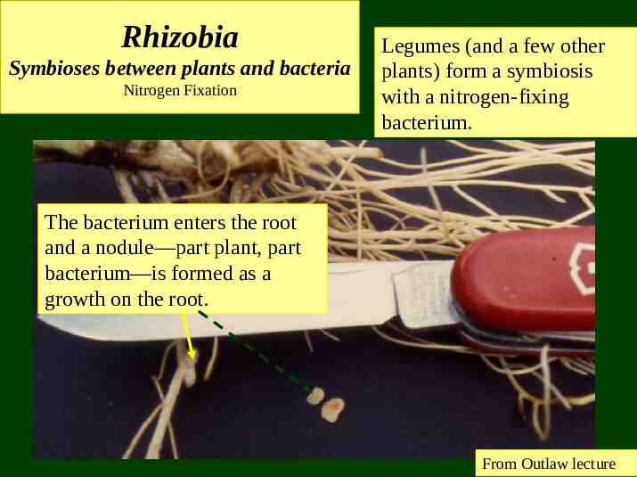
Rhizobia Symbioses between plants and bacteria Nitrogen Fixation Legumes (and a few other plants) form a symbiosis with a nitrogen-fixing bacterium. The bacterium enters the root and a nodule—part plant, part bacterium—is formed as a growth on the root. From Outlaw lecture
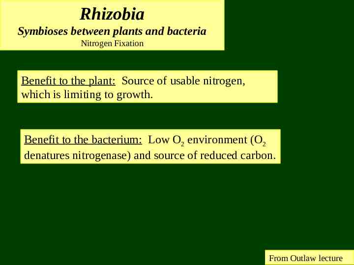
Rhizobia Symbioses between plants and bacteria Nitrogen Fixation Benefit to the plant: Source of usable nitrogen, which is limiting to growth. Benefit to the bacterium: Low O2 environment (O2 denatures nitrogenase) and source of reduced carbon. From Outlaw lecture
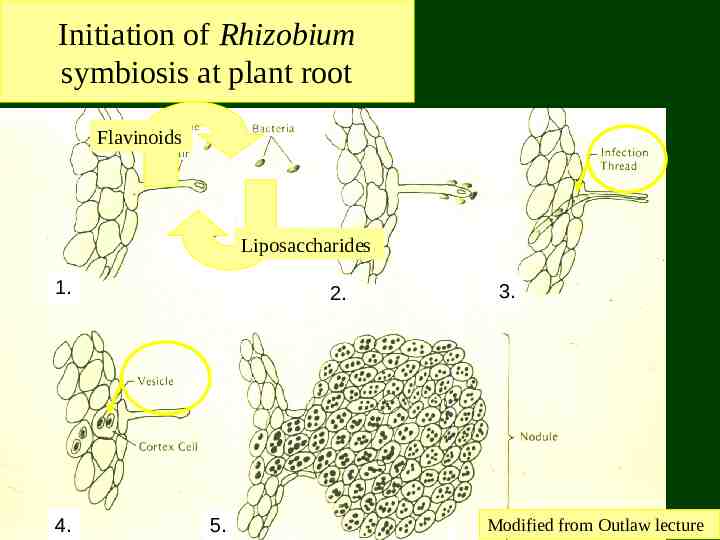
Initiation of Rhizobium symbiosis at plant root Flavinoids Liposaccharides 1. 4. 2. 5. 3. Modified from Outlaw lecture
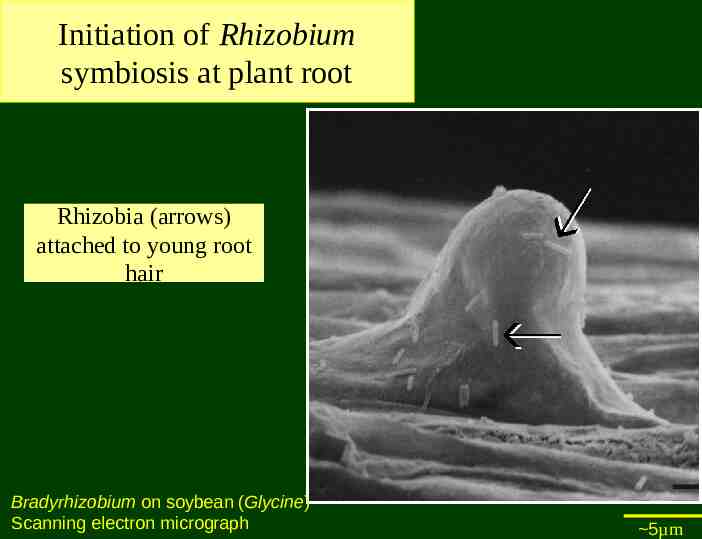
Initiation of Rhizobium symbiosis at plant root Rhizobia (arrows) attached to young root hair Bradyrhizobium on soybean (Glycine) Scanning electron micrograph 5µm
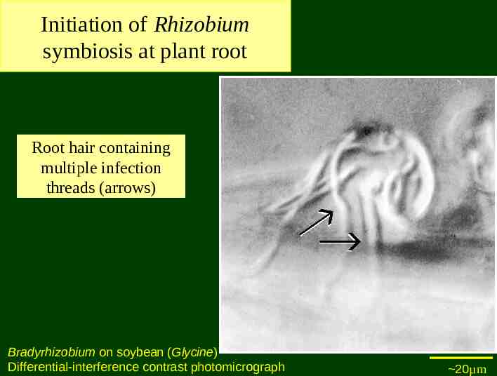
Initiation of Rhizobium symbiosis at plant root Root hair containing multiple infection threads (arrows) Bradyrhizobium on soybean (Glycine) Differential-interference contrast photomicrograph 20µm
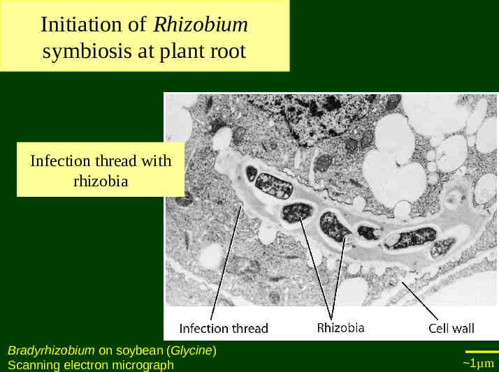
Initiation of Rhizobium symbiosis at plant root Infection thread with rhizobia Bradyrhizobium on soybean (Glycine) Scanning electron micrograph 1µm
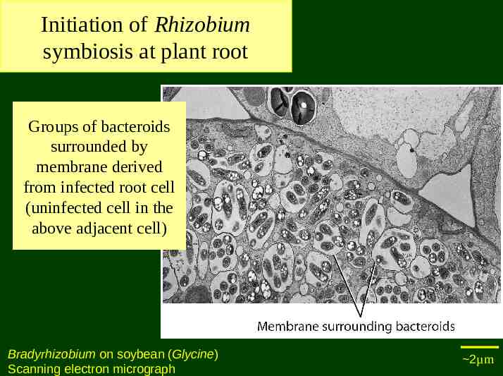
Initiation of Rhizobium symbiosis at plant root Groups of bacteroids surrounded by membrane derived from infected root cell (uninfected cell in the above adjacent cell) Bradyrhizobium on soybean (Glycine) Scanning electron micrograph 2µm
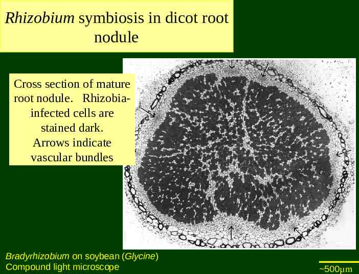
Rhizobium symbiosis in dicot root nodule Cross section of mature root nodule. Rhizobiainfected cells are stained dark. Arrows indicate vascular bundles Bradyrhizobium on soybean (Glycine) Compound light microscope 500µm
A few examples Flowering plants and pollinating animals Humans and domesticated plants and animals Humans and bacteria in their digestive system Endosymbionts Origination of mitochondria and chloroplasts Plants and bacteria Rhizobia (also an example of endosymbiosis) Humans and fungi Leafcutter ants and fungi Lichens
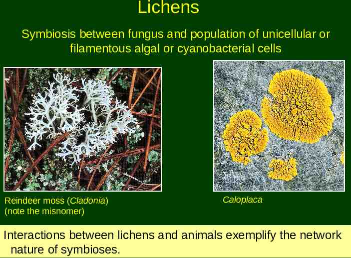
Lichens Symbiosis between fungus and population of unicellular or filamentous algal or cyanobacterial cells Reindeer moss (Cladonia) (note the misnomer) Caloplaca Interactions between lichens and animals exemplify the network nature of symbioses.
A few examples Flowering plants and pollinating animals Humans and domesticated plants and animals Humans and bacteria in their digestive system Endosymbionts Origination of mitochondria and chloroplasts Plants and bacteria Rhizobia (also an example of endosymbiosis) Humans and fungi Leafcutter ants and fungi Lichens Plants and fungi Mycorrhizae
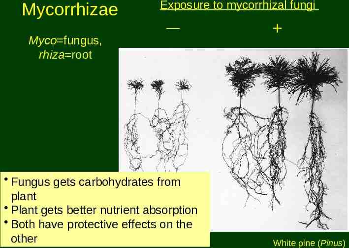
Mycorrhizae Exposure to mycorrhizal fungi Myco fungus, rhiza root Fungus gets carbohydrates from plant Plant gets better nutrient absorption Both have protective effects on the other White pine (Pinus)
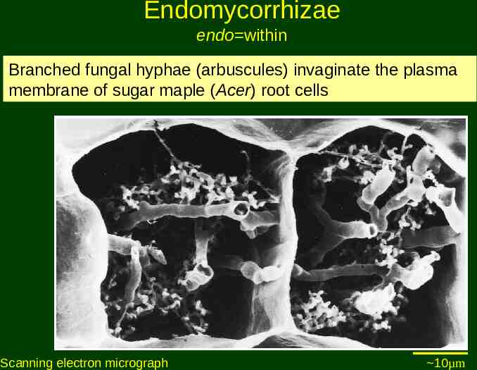
Endomycorrhizae endo within Branched fungal hyphae (arbuscules) invaginate the plasma membrane of sugar maple (Acer) root cells Scanning electron micrograph 10µm
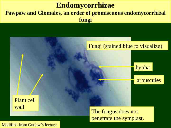
Endomycorrhizae Pawpaw and Glomales, an order of promiscuous endomycorrhizal fungi Fungi (stained blue to visualize) hypha arbuscules Plant cell wall Modified from Outlaw’s lecture The fungus does not penetrate the symplast.
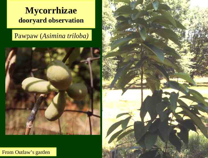
Mycorrhizae dooryard observation Pawpaw (Asimina triloba) From Outlaw’s garden
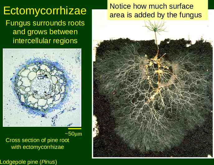
Ectomycorrhizae Fungus surrounds roots and grows between intercellular regions 50µm Cross section of pine root with ectomycorrhizae Lodgepole pine (Pinus) Notice how much surface area is added by the fungus
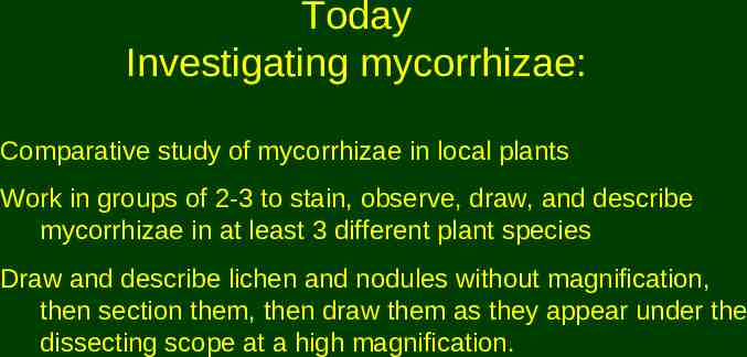
Today Investigating mycorrhizae: Comparative study of mycorrhizae in local plants Work in groups of 2-3 to stain, observe, draw, and describe mycorrhizae in at least 3 different plant species Draw and describe lichen and nodules without magnification, then section them, then draw them as they appear under the dissecting scope at a high magnification.