Systemic Lupus Erythematosus Steve Beesley

19 Slides871.50 KB
Systemic Lupus Erythematosus Steve Beesley
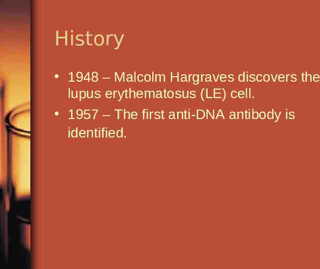
History 1948 – Malcolm Hargraves discovers the lupus erythematosus (LE) cell. 1957 – The first anti-DNA antibody is identified.
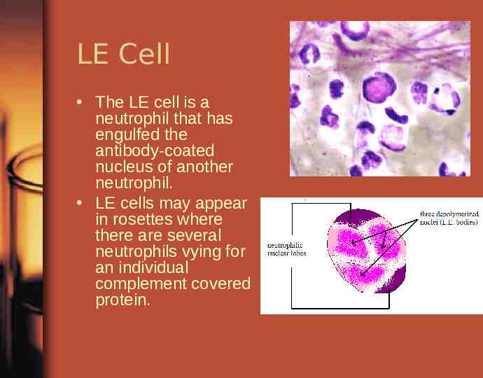
LE Cell The LE cell is a neutrophil that has engulfed the antibody-coated nucleus of another neutrophil. LE cells may appear in rosettes where there are several neutrophils vying for an individual complement covered protein.
Genetic Associations HLA’s are loci on genes that code for certain β chain on the MHC complex HLA-DR2 HLA-DR3 HLA-DQB1 – Involved in mediating production of antibodies to ds-DNA
Symptoms Non-specific: – – – – – – Fatigue Weight loss Malaise generally feeling ill Fever Anorexia (over time) Arthritis 90% of patients experience arthritic symptoms Symmetrical Appears in hands, wrists, and knees mainly
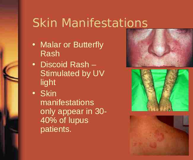
Skin Manifestations Malar or Butterfly Rash Discoid Rash – Stimulated by UV light Skin manifestations only appear in 3040% of lupus patients.
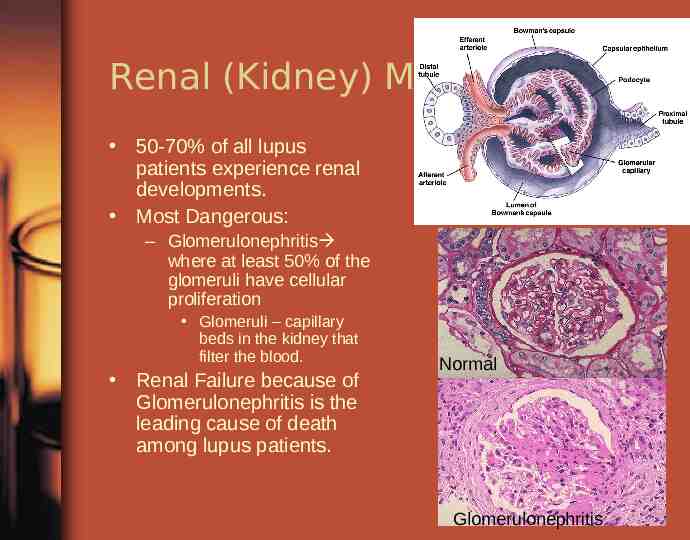
Renal (Kidney) Manifestations 50-70% of all lupus patients experience renal developments. Most Dangerous: – Glomerulonephritis where at least 50% of the glomeruli have cellular proliferation Glomeruli – capillary beds in the kidney that filter the blood. Renal Failure because of Glomerulonephritis is the leading cause of death among lupus patients. Normal Glomerulonephritis
Other Manifestations Cardiac Central Nervous System Hematological
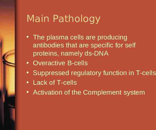
Main Pathology The plasma cells are producing antibodies that are specific for self proteins, namely ds-DNA Overactive B-cells Suppressed regulatory function in T-cells Lack of T-cells Activation of the Complement system
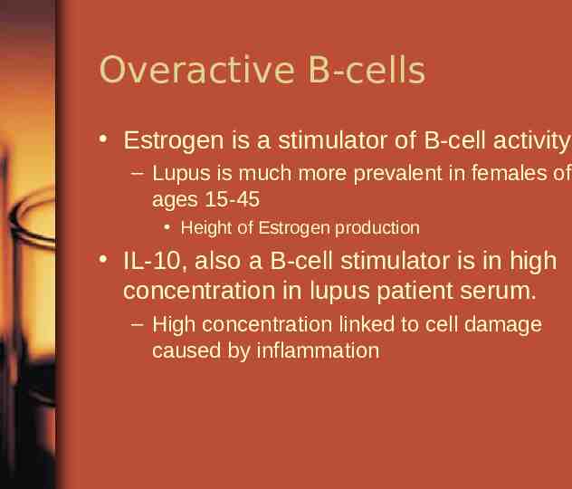
Overactive B-cells Estrogen is a stimulator of B-cell activity – Lupus is much more prevalent in females of ages 15-45 Height of Estrogen production IL-10, also a B-cell stimulator is in high concentration in lupus patient serum. – High concentration linked to cell damage caused by inflammation
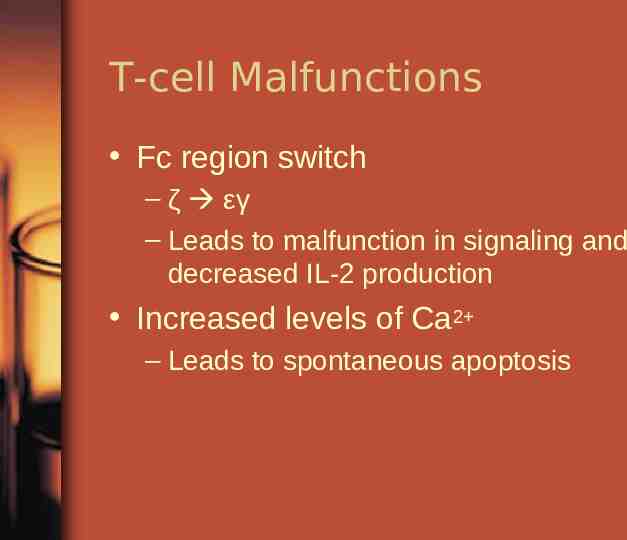
T-cell Malfunctions Fc region switch – ζ εγ – Leads to malfunction in signaling and decreased IL-2 production Increased levels of Ca2 – Leads to spontaneous apoptosis
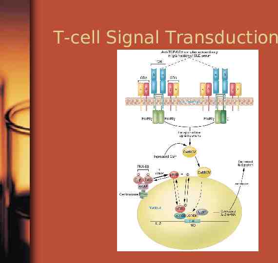
T-cell Signal Transduction
Activation of Complement System Complement system is activated by the binding of antibodies to foreign debris. – In this case its over activation RBCs lack CR1 receptor – Decreasing the affective removal of complexes
IgG Pathogen IgG is the most “pathogenic” because it forms intermediate sized complexes that can get to the small places and block them.
DNA is the Main man DNA is the main antigen for which antibodies are formed. Extracellular DNA has an affinity for basement membrane where it is bound by autoantibodies. Classical thickening of the basement membrane
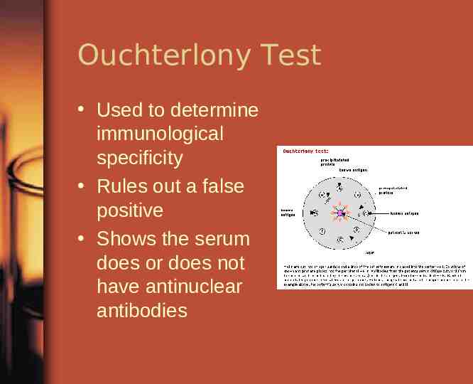
Testing ESR Urinalysis Complement Test – Tests levels of C3, C4, CH50 – Low levels indicates possible presence of disease FANA – Fluorescent antinuclear antibody Ouchterlony Test – shows interactions
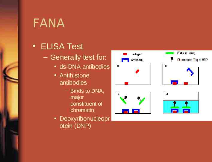
FANA ELISA Test – Generally test for: ds-DNA antibodies Antihistone antibodies – Binds to DNA, major constituent of chromatin Deoxyribonucleopr otein (DNP)
Ouchterlony Test Used to determine immunological specificity Rules out a false positive Shows the serum does or does not have antinuclear antibodies
Summary Lupus Autoimmunity – Systemic and affects connective tissue Caused by malfunctions of: – – – – T-cells B-cells Complement System Signal Transduction Can be lethal or not Unique to each individual