RSNA 2008 – Course 1029 Electronic Reports: HL7 CDA (Clinical
52 Slides3.08 MB
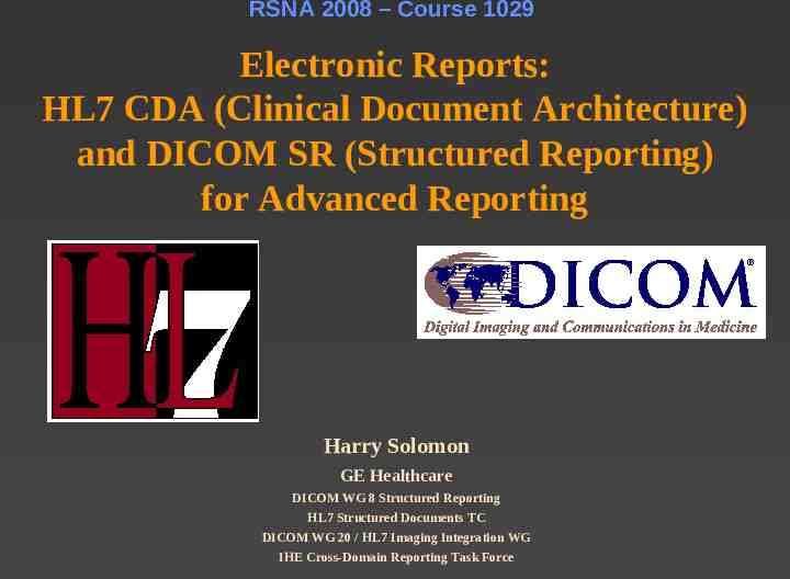
RSNA 2008 – Course 1029 Electronic Reports: HL7 CDA (Clinical Document Architecture) and DICOM SR (Structured Reporting) for Advanced Reporting Harry Solomon GE Healthcare DICOM WG 8 Structured Reporting HL7 Structured Documents TC DICOM WG 20 / HL7 Imaging Integration WG IHE Cross-Domain Reporting Task Force
Disclosure Harry Solomon – Employee, GE Healthcare – Instructor, Medical Informatics, Northwestern University 2
Acknowledgement Fred Behlen, s co-author of a previous version of this presentation Fred Behlen, Bob Dolin, Liora Alschuler, Calvin Beebe – cochairs of HL7 Structured Documents Technical Committee, and authors of presentations on CDA used in this talk Dave Clunie – former co-chair of DICOM Standards Committee, and author of the definitive book on DICOM Structured Reporting Kevin O’Donnell – IHE Reporting Task Force 3
Objective s the key elements for effective radiology Understand reporting, and issues with electronic reporting workflows Understand the uses of HL7 CDA (Clinical Document Architecture) and DICOM SR (Structured Reporting) for advanced reporting workflows 4
Is this an electronic report? MSH \& RIS GOOD HOSPITAL 198808181126 ORU O01 ORU O01 P 2.6 cr PID 1 PATID1234 5 M11 ADT1 MR GOOD HOSPITAL 123456789 USSSA SS EVERYMAN ADAM A III 19610615 M C 2222 HOME STREET GREENSBORO NC 27401‑1020 GL (555) 555‑2004 S PATID12345001 2 M10 ADT1 AN A 9 87654 NC cr PV1 1 I 2000 2012 01 004777 ATTEND AARON A SUR ADM A0 cr OBR 1 P8754 OE XR1501 XR 24646-2 CXR PA LAT LN 198703290800 4010 INTERN IRVING I MD L . cr OBX 1 TX 24646-2 CXR PA LAT LN Infiltrate probably representing bronchopneumonia in the right lower lobe. Also pulmonary venous congestion cardiomegaly and cephalization, indicating early congestive heart failure. Followup CXR 1 month. . cr 5
For our purposes An electronic report is created using computer based techniques (workflow), includes some amount of structured and coded content, and may include “multimedia” (for radiology, images) We will look at two technology standards that apply to electronic reporting 6
Key Elements of Radiology Reporting 7
Paper or Electronic Accurately convey the findings to the referring physician Reports – Reflect the competence of the radiologist Timely communication for patient care Archived in the patient medical record Legal record of imaging exam – Radiologist signature Support ‘secondary’ uses – – – – Charge capture and billing Teaching and research Clinical data registries, clinical trials Process improvement Produced making best use of radiologist’s time 8 Typical busy radiologist at Northwestern Memorial Hospital
Benefits ( ) and challenges (-) Accuracy of Electronic Reports Drive for quality improvement with quantitative data, CAD and other measurements Possible major benefit with attached key images and graphical analysis (picture 1000 words) – Will systems support graphical reports? Timely communication Probable improvement Archived in the patient medical record – Where is the electronic medical record? (distributed, multiple copies) 9
Benefits and challenges of Electronic Reports Legal record (cont’d) – What is a valid electronic signature? – Is an exact visual reproduction required, or only exact semantic content? Secondary uses Huge potential improvement, especially with structured and coded data More accurate billing (avoid undercoding) Use of radiologist’s time – Potential negative impact with transition from traditional dictation workflow – Radiologist pays the cost for improvements downstream 10
Planning for electronic reporting What are your goals ? – Better capture of sonographer measurements into report – Add key images into reports – Ability to do research / data mining What kinds of reports do you need? – – – – – Text only Text image references Structured text Structured text coded content Multimedia 11
This is Process Reengineering! Transition to electronic reports is hard – – – – New systems New architectures New policies and procedures Organizationally disjunct costs/benefits Minimize the risk and the effort – A standards-based approach – Incremental evolution from current workflow – Leverage the work of IHE (Integrating the Healthcare Enterprise) 12
Radiology Reporting Workflows 13
Reporting Starts Before the Radiologist Sees the Study Reason for exam (from order), patient history Technical aspects of procedure – Protocol – Exam notes from tech Post-processing results – Measurement and analysis applications (e.g., vascular, obstetric, cardiac) by tech – Computer Aided Detection results – Produced on modality, imaging workstation, or CAD server These need to get to the radiologist and integrated into the report 14
Reporting Integration Review(1) study evidence – Order and relevant clinical information – Images and relevant priors – Tech notes and post-processing results Radiologist interpretation – on imaging workstation – Annotation (virtual grease pencil) – Key image selection – Measurement and analysis applications by radiologist Radiologist findings reporting – on a different system? – Dictation transcription / speech recognition – Structured data entry (forms-based) 15 Where’s Waldo going to prepare his report?
Reporting Integration Report(2) assembly – Findings and selected evidence/interpretation results Radiologist signature – Auditable action, or digital encryption-based Report communication – To referring physician – To “secondary” users (billing!, quality improvement) Report archive – And subsequent access 16
Diagnostic Image Viewing reporting Application User control Reporting Application Diagnostic report * **** * **** ** ** ** ** * ***** * **** * ***** ***** * **** * **** ** ** **** * ***** ***** ***** * **** * U NIVE R SITY OF CH I CAGO HOSP IT AL S R **** A DIOLO GY ** CO ** N SULT * *** *** * ***** ***** ***** * ** *** ****A*TION ***** ***** ***** * ***** * **** ** *** ** *** ** 3 42 0 2 /05/ 96 UNIV ER SIT Y OF CHI C AGO H OSPIT ALS B HIS # : 12 34 56 7 INPA T IENT 201- 2 3-90 RA DIO LO GY CON S ULTAT ION H emat3o42 logy O nc ol o gy 02//0 5/96 CH ANDLE R, CA R OLYN M itchBeHIS ll-6#: NE 12345 67 INP A TIEN T H ema tol o gy / Oncol ogy A dmitMtitc inghel Dilag no si s : NE U TROP E NIC F EVER; HYPE R BILI RU BE MIA -6NE C lini c al d at a: Bil iar y t ube c heck. CAadmi rl tti M. G er s, M D s: n omp g Dia gnosi 49 F EMALE 201 -2 3-9 0 C H ANDL ER , C AR OLY N 49 FE MAL E N E UTRO PE NIC F EV ER; H YPERB ILIRU BEMIA C lin ica l data : Bil iar y t ube ch ec k. C hang e PerCar c l Bi M. li ary Drrain G ompe s, age MD C ath Pr oce d -- C han ge P erc B ili ar y Dra i nage Cat h Pro c ed CO M PARI SO N: 0 7/ 2 3/95 a nd 0 6 /27/9 5 Exa m #4 6 on 01/ 08 /96 -- Exam #46 o n 01/ 0 8/96 FI N DING S:A RISON A ft er : the r oced and u re 06 wa /27 s exp C OMP 07/ p 23/95 /9 5l aine d to th e pati e nt an d inf ormed -- Ex am #4 7 on 0 2/05/ 9 6 & In t F IND I NGS: After the p roce du re wa s expl a ined to th e pat i ent a n d in fo rme d & Int -- Ex am #4 7 on 0 2/05 /9 6 FI N DING S: A s ab o ve. IM P RESS IO N: F IND I NGS: As ab ove. Su c cess fuElSSION bil ia:ry t ub e ch ange, and f indi ng s c ons is ten t with inte r val t umor I MPR g rowt h . S ucc e ssful bili a ry t u be c h ange , and fi nd ings cons is ten t with inte r val tu mor Sigmrow on th. A . T emp la r, M D / R ic hard Nixo n, MD (R1 9) Si g ned 02 /9 /9 6 a t 8:4 8 AM 3 S imo n A. T empl a r, MD / R i char d Nix on , M D (R 19) S ign e d 02/ 9/96 at 8: 4 8 AM 3 Diagnostic Images Image Sources Viewing settings (ww/wl, rotation/flip) PACS Archive Orders, Prior Reports Report Information System 17
Reporting with annotation Image Viewing (use case desired) Application User control Diagnostic Images Image Sources Viewing settings (ww/wl) PACS Archive Reporting Application Diagnostic report Image references & annotation Orders, Prior Reports * **** * **** ** ** ** ** * ***** * **** * ***** ***** * **** * **** ** ** **** * ***** ***** ***** * **** * U NIVE R SITY OF CH I CAGO HOSP IT AL S R **** A DIOLO GY ** CO ** N SULT * *** *** * ***** ***** ***** * ** *** ****A*TION ***** ***** ***** * ***** * **** ** *** ** *** ** 3 42 0 2 /05/ 96 UNIV ER SIT Y OF CHI C AGO H OSPIT ALS B HIS # : 12 34 56 7 INPA T IENT 201- 2 3-90 RA DIO LO GY CON S ULTAT ION H emat3o42 logy O nc ol o gy 02//0 5/96 CH ANDLE R, CA R OLYN M itchBeHIS ll-6#: NE 12345 67 INP A TIEN T H ema tol o gy / Oncol ogy A dmitMtitc inghel Dilag no si s : NE U TROP E NIC F EVER; HYPE R BILI RU BE MIA -6NE C lini c al d at a: Bil iar y t ube c heck. CAadmi rl tti M. G er s, M D s: n omp g Dia gnosi 49 F EMALE 201 -2 3-9 0 C H ANDL ER , C AR OLY N 49 FE MAL E N E UTRO PE NIC F EV ER; H YPERB ILIRU BEMIA C lin ica l data : Bil iar y t ube ch ec k. C hang e PerCar c l Bi M. li ary Drrain G ompe s, age MD C ath Pr oce d -- C han ge P erc B ili ar y Dra i nage Cat h Pro c ed CO M PARI SO N: 0 7/ 2 3/95 a nd 0 6 /27/9 5 Exa m #4 6 on 01/ 08 /96 -- Exam #46 o n 01/ 0 8/96 FI N DING S:A RISON A ft er : the r oced and u re 06 wa /27 s exp C OMP 07/ p 23/95 /9 5l aine d to th e pati e nt an d inf ormed -- Ex am #4 7 on 0 2/05/ 9 6 & In t F IND I NGS: After the p roce du re wa s expl a ined to th e pat i ent a n d in fo rme d & Int -- Ex am #4 7 on 0 2/05 /9 6 FI N DING S: A s ab o ve. IM P RESS IO N: F IND I NGS: As ab ove. Su c cess fuElSSION bil ia:ry t ub e ch ange, and f indi ng s c ons is ten t with inte r val t umor I MPR g rowt h . S ucc e ssful bili a ry t u be c h ange , and fi nd ings cons is ten t with inte r val tu mor Sigmrow on th. A . T emp la r, M D / R ic hard Nixo n, MD (R1 9) Si g ned 02 /9 /9 6 a t 8:4 8 AM 3 S imo n A. T empl a r, MD / R i char d Nix on , M D (R 19) S ign e d 02/ 9/96 at 8: 4 8 AM 3 Report with image references & annotation Information System 18
Reporting with annotation Image Viewing (what’s available) Application User control Image references & annotation Diagnostic Images Image Sources Viewing settings, image references & annotation PACS Archive Reporting Application Diagnostic report * **** * **** ** ** ** ** * ***** * **** * ***** ***** * **** * **** ** ** **** * ***** ***** ***** * **** * U NIVE R SITY OF CH I CAGO HOSP IT AL S R **** A DIOLO GY ** CO ** N SULT * *** *** * ***** ***** ***** * ** *** ****A*TION ***** ***** ***** * ***** * **** ** *** ** *** ** 3 42 0 2 /05/ 96 UNIV ER SIT Y OF CHI C AGO H OSPIT ALS B HIS # : 12 34 56 7 INPA T IENT 201- 2 3-90 RA DIO LO GY CON S ULTAT ION H emat3o42 logy O nc ol o gy 02//0 5/96 CH ANDLE R, CA R OLYN M itchBeHIS ll-6#: NE 12345 67 INP A TIEN T H ema tol o gy / Oncol ogy A dmitMtitc inghel Dilag no si s : NE U TROP E NIC F EVER; HYPE R BILI RU BE MIA -6NE C lini c al d at a: Bil iar y t ube c heck. CAadmi rl tti M. G er s, M D s: n omp g Dia gnosi 49 F EMALE 201 -2 3-9 0 C H ANDL ER , C AR OLY N 49 FE MAL E N E UTRO PE NIC F EV ER; H YPERB ILIRU BEMIA C lin ica l data : Bil iar y t ube ch ec k. C hang e PerCar c l Bi M. li ary Drrain G ompe s, age MD C ath Pr oce d -- C han ge P erc B ili ar y Dra i nage Cat h Pro c ed CO M PARI SO N: 0 7/ 2 3/95 a nd 0 6 /27/9 5 Exa m #4 6 on 01/ 08 /96 -- Exam #46 o n 01/ 0 8/96 FI N DING S:A RISON A ft er : the r oced and u re 06 wa /27 s exp C OMP 07/ p 23/95 /9 5l aine d to th e pati e nt an d inf ormed -- Ex am #4 7 on 0 2/05/ 9 6 & In t F IND I NGS: After the p roce du re wa s expl a ined to th e pat i ent a n d in fo rme d & Int -- Ex am #4 7 on 0 2/05 /9 6 FI N DING S: A s ab o ve. IM P RESS IO N: F IND I NGS: As ab ove. Su c cess fuElSSION bil ia:ry t ub e ch ange, and f indi ng s c ons is ten t with inte r val t umor I MPR g rowt h . S ucc e ssful bili a ry t u be c h ange , and fi nd ings cons is ten t with inte r val tu mor Sigmrow on th. A . T emp la r, M D / R ic hard Nixo n, MD (R1 9) Si g ned 02 /9 /9 6 a t 8:4 8 AM 3 S imo n A. T empl a r, MD / R i char d Nix on , M D (R 19) S ign e d 02/ 9/96 at 8: 4 8 AM 3 Orders, Prior Reports Report Information System 19
Reporting with Image Viewing measurements Application User control Reporting Application Diagnostic report * **** * **** ** ** ** ** * ***** * **** * ***** ***** * **** * **** ** ** **** * ***** ***** ***** * **** * U NIVE R SITY OF CH I CAGO HOSP IT AL S R **** A DIOLO GY ** CO ** N SULT * *** *** * ***** ***** ***** * ** *** ****A*TION ***** ***** ***** * ***** * **** ** *** ** *** ** 3 42 0 2 /05/ 96 UNIV ER SIT Y OF CHI C AGO H OSPIT ALS B HIS # : 12 34 56 7 INPA T IENT 201- 2 3-90 RA DIO LO GY CON S ULTAT ION H emat3o42 logy O nc ol o gy 02//0 5/96 CH ANDLE R, CA R OLYN M itchBeHIS ll-6#: NE 12345 67 INP A TIEN T H ema tol o gy / Oncol ogy A dmitMtitc inghel Dilag no si s : NE U TROP E NIC F EVER; HYPE R BILI RU BE MIA -6NE C lini c al d at a: Bil iar y t ube c heck. CAadmi rl tti M. G er s, M D s: n omp g Dia gnosi 49 F EMALE 201 -2 3-9 0 C H ANDL ER , C AR OLY N 49 FE MAL E N E UTRO PE NIC F EV ER; H YPERB ILIRU BEMIA C lin ica l data : Bil iar y t ube ch ec k. C hang e PerCar c l Bi M. li ary Drrain G ompe s, age MD C ath Pr oce d -- C han ge P erc B ili ar y Dra i nage Cat h Pro c ed CO M PARI SO N: 0 7/ 2 3/95 a nd 0 6 /27/9 5 Exa m #4 6 on 01/ 08 /96 -- Exam #46 o n 01/ 0 8/96 FI N DING S:A RISON A ft er : the r oced and u re 06 wa /27 s exp C OMP 07/ p 23/95 /9 5l aine d to th e pati e nt an d inf ormed -- Ex am #4 7 on 0 2/05/ 9 6 & In t F IND I NGS: After the p roce du re wa s expl a ined to th e pat i ent a n d in fo rme d & Int -- Ex am #4 7 on 0 2/05 /9 6 FI N DING S: A s ab o ve. IM P RESS IO N: F IND I NGS: As ab ove. Su c cess fuElSSION bil ia:ry t ub e ch ange, and f indi ng s c ons is ten t with inte r val t umor I MPR g rowt h . S ucc e ssful bili a ry t u be c h ange , and fi nd ings cons is ten t with inte r val tu mor Sigmrow on th. A . T emp la r, M D / R ic hard Nixo n, MD (R1 9) Si g ned 02 /9 /9 6 a t 8:4 8 AM 3 S imo n A. T empl a r, MD / R i char d Nix on , M D (R 19) S ign e d 02/ 9/96 at 8: 4 8 AM 3 EDD 0921 BPD 5.2 cm Diagnostic images & measurements Image Sources Viewing settings & confirmed measurements PACS Archive Measurement Sources Orders, Prior Reports Report Information System 20
The issues How do we bridge the gap between the imaging side and the reporting side – Annotations, key images, and measurements How do we include these enhanced features in reports? 21
HL7 Clinical Document Architecture Overview HL7 is a Standards Development Organization whose domain is clinical and administrative data 22
HL7 Clinical Document Architecture The scope of the CDA is the standardization of clinical documents for exchange. A clinical document is a record of observations and other services with the following characteristics: – – – – – Persistence Stewardship Potential for authentication Wholeness Human readability A CDA document is a defined and complete information object that can exist outside of a message, and can include text, images, sounds, and other multimedia content. 23
Why do you need to know about Executive Order 13,410 and EHR Safe Harbors CDA? Provision (Stark Act relaxation): certain healthcare IT systems must comply with federally recognized interoperability specifications January 2008: HHS Secretary Leavitt recognizes first HITSP* Interoperability Specifications, including several components using CDA While not (yet) specified for interoperability of radiology reports, HITSP considers CDA as basis for clinical documentation going forward *Healthcare IT Standards Panel of American National Standards Institute (ANSI), tasked 24 by Dept of Health & Human Services to recommend harmonized standards
Clinical Document Characteristics Persistence – Documents exist over time and can be used in many contexts Stewardship – Documents must be managed, shared by the steward Potential for authentication – Intended use as medico-legal documentation Wholeness – Document includes its relevant context Human readability – Essential for human authentication 25
CDA Use Cases Diagnostic and therapeutic procedure reports Encounter / discharge summaries Patient history & physical Referrals Claims attachments Consistent format for all clinical documents 26
Key Aspects of the CDA documents CDA are encoded in Extensible Markup Language (XML) CDA documents derive their meaning from the HL7 v3 Reference Information Model (RIM ) and use HL7 v3 Data Types A CDA document consists of a header and a body – Header is consistent across all clinical documents - identifies and classifies the document, provides information on patient, provider, encounter, and authentication – Body contains narrative text / multimedia content (level 1), optionally augmented by coded equivalents (levels 2 & 3) 27
CDA Release Standard 1 (2000) – Standalone standard, based on early draft v3 RIM – Level 1 narrative and multimedia Release 2 (2005) – Incorporated into HL7 v3 Standard (Normative Edition) – Level 2 structured narrative and multimedia, plus Level 3 coded statements Implementation Guides – – – – – HL7 Care Record Summary (CRS) ASTM/HL7 Continuity of Care Document (CCD) IHE Patient Care Coordination Templates Common Document Types project (CDA4CDT) HL7 Diagnostic Imaging Report Implementation Guide New 28
CDA Release 2 Information Model Header Participants Start Here Doc ID &Type Body Context Sections/ Headings Clinical Statements/ Coded Entries Extl 29 Refs
CDA Structured Body Arrows are Act Relationships Has component, Derived from, etc. Entries are coded clinical statements Observation, Procedure, Substance administration, etc. Structured Body Section Text Section Text Section Text Section Text Section Text Entry Coded statement Section Text Entry Coded statement Entry Coded statement 30
Sam ple CD A 31
Principle of Human Readability: Narrative and Coded Information CDA structured body requires human-readable “Narrative Block”, all that is needed to reproduce the legally attested clinical content CDA allows optional machine-readable coded “Entries”, which drive automated processes By starting with a base of text, CDA allows incremental improvement to amount of coded data without breaking the model 32
Narrative and Coded Entry Example 33
CDA Non-XML Body Alternative to XML Structured Body Standard CDA header “wraps” existing document – Allows document management with consistent metadata Body can be any MIME* type – Especially PDF (IHE Scanned Document Profile) *Multi-part Internet Mail Extension 34
CD Published by HL7 A – Care Record Summary – encounter notes, discharge summary – Continuity of Care Document – transfer of care (harmonized with Imp ASTM Continuity of Care Record) – Diagnostic Imaging Report – with robust references to DICOM objects lem Published by IHE Patient Care Coordination enta – Emergency Department Referral – Pre-procedure History and Physical tion – Scanned Documents – Personal Health Record Extract – BasicGui Patient Privacy Consents – Antepartum Summary desDepartment Encounter Summary – Emergency 35
Header Diagnostic Imaging Report Implementation Guide Structured Body Section DICOM Object Catalog Section Reason for Study Section Findings Section Patient History Entries DICOM Study, Series, Image References References to DICOM objects in hierarchical context using native DICOM or WADO access References to DICOM images with optional Presentation State annotations Section Impressions Section Procedure Description Section Comparison Study Section Recommendations Entries (Annotated) Image References Section Key Images 36
DICOM Structured Reporting Overview DICOM is a Standards Development Organization whose domain is biomedical imaging 37
DICOM Structured Reporting The scope of DICOM SR is the standardization of documents in the imaging environment. SR documents record observations made for an imaging-based diagnostic or interventional procedure, particularly those that describe or reference images, waveforms, or specific regions of interest. 38
Why do you need to know about DICOM DICOMSR? SR is used in key subspecialty areas that produce structured data in the course of image acquisition or post-processing, where: – Leveraging the DICOM infrastructure is easy and desirable – Results should be managed with other study evidence Examples – – – – – Sonographer measurements Computer-aided detection results QC notes about images Radiation dose reports Image exchange manifests 39
Key Aspects of DICOM SR documents SR are encoded using DICOM standard data elements and leverage DICOM network services (storage, query/retrieve) SR uses DICOM Patient/Study/Series information model (header), plus hierarchical tree of “Content Items” Extensive mandatory use of coded content – Allows use of vocabulary/codes from non-DICOM sources Templates define content constraints for specific types of documents / reports 40
SR Content Item Tree Arrows are parent-child relationships Contains, Has properties, Inferred from, etc. Content Items are units of meaning Text, Numeric, Code, Image, Spatial coordinates, etc. Root Content Item Document Title Content Item Content Item Content Item Content Item Content Item Content Item Content Item Content Item Content Item 41
DICOM SR Example 42
DICOM SR Object Enhanced and Comprehensive - Text, coded content, numeric Classes measurements, spatial and temporal ROI references – Templates for ultrasound, cardiac imaging CAD - Automated analysis results (mammo, chest, colon) Key Object Selection (KO) - Flags one or more images – Purpose (for referring physician, for surgery ) and textual note – Used for key image notes and image manifests (in IHE profiles) Procedure Log - For extended duration procedures (e.g., cath) Radiation Dose Report - Projection X-ray; CT 43
HL7 CDA and DICOM SR Compare and Contrast and Collaborate 44
“Evidence” and Evidence Documents “Reports” – Includes measurements, procedure logs, CAD results, etc., created in the imaging context, and together with images are interpreted by a radiologist to produce a report – The radiologist may quote or copy parts of Evidence Documents into the report, but doing so is part of the interpretation process at his discretion – Appropriate to be stored in PACS as DICOM SR objects, with same (legal/distribution) status as images Reports – Become part of the patient’s medical record, with potentially wide distribution – Good match to HL7 CDA 45
DICOM-HL7 Synergy DICOM(1) and HL7 have recognized the need to work together DICOM SR and HL7 CDA are congruent in key areas – – – – Document persistence Document identification, versioning and type code Document’s relation to the patient and to the authoring physicians Coded content using external vocabularies SR strength in robust image-related semantic content; CDA strength in human readable narrative report 46
DICOM-HL7 Synergy Methods(2) for referencing CDA documents from within DICOM objects, and vice versa CDA documents can be included on DICOM exchange disks – As native CDA files, or encapsulated in a DICOM file – Indexed in DICOMDIR for integration with DICOM applications Transcoding from SR to CDA feasible for measurements, image references, observations DICOM WG10 (Strategic Advisory) suggested composing radiology reports directly in CDA format when appropriate 47
Approaches to integration Use these standards! Ask for them from your IT providers Leverage them in new combinations to achieve desired electronic reporting capabilities Evolve from current workflows – but recognize there may be process reengineering 48
Loosely integrated reporting – Image Viewing Reporting add key images to reports Application Application User control Image references & annotation Viewing settings, Diagnostic image references Images & annotation Image references Image PACS & annotation Sources Archive Image retrieval Diagnostic report * **** * **** ** ** ** ** * ***** * **** * ***** ***** * **** * **** ** ** **** * ***** ***** ***** * **** * U NIVE R SITY OF CH I CAGO HOSP IT AL S R **** A DIOLO GY ** CO ** N SULT * *** *** * ***** ***** ***** * ** *** ****A*TION ***** ***** ***** * ***** * **** ** *** ** *** ** 3 42 0 2 /05/ 96 UNIV ER SIT Y OF CHI C AGO H OSPIT ALS B HIS # : 12 34 56 7 INPA T IENT 201- 2 3-90 RA DIO LO GY CON S ULTAT ION H emat3o42 logy O nc ol o gy 02//0 5/96 CH ANDLE R, CA R OLYN M itchBeHIS ll-6#: NE 12345 67 INP A TIEN T H ema tol o gy / Oncol ogy A dmitMtitc inghel Dilag no si s : NE U TROP E NIC F EVER; HYPE R BILI RU BE MIA -6NE C lini c al d at a: Bil iar y t ube c heck. CAadmi rl tti M. G er s, M D s: n omp g Dia gnosi 49 F EMALE 201 -2 3-9 0 C H ANDL ER , C AR OLY N 49 FE MAL E N E UTRO PE NIC F EV ER; H YPERB ILIRU BEMIA C lin ica l data : Bil iar y t ube ch ec k. C hang e PerCar c l Bi M. li ary Drrain G ompe s, age MD C ath Pr oce d -- C han ge P erc B ili ar y Dra i nage Cat h Pro c ed CO M PARI SO N: 0 7/ 2 3/95 a nd 0 6 /27/9 5 Exa m #4 6 on 01/ 08 /96 -- Exam #46 o n 01/ 0 8/96 FI N DING S:A RISON A ft er : the r oced and u re 06 wa /27 s exp C OMP 07/ p 23/95 /9 5l aine d to th e pati e nt an d inf ormed -- Ex am #4 7 on 0 2/05/ 9 6 & In t F IND I NGS: After the p roce du re wa s expl a ined to th e pat i ent a n d in fo rme d & Int -- Ex am #4 7 on 0 2/05 /9 6 FI N DING S: A s ab o ve. IM P RESS IO N: F IND I NGS: As ab ove. Su c cess fuElSSION bil ia:ry t ub e ch ange, and f indi ng s c ons is ten t with inte r val t umor I MPR g rowt h . S ucc e ssful bili a ry t u be c h ange , and fi nd ings cons is ten t with inte r val tu mor Sigmrow on th. A . T emp la r, M D / R ic hard Nixo n, MD (R1 9) Si g ned 02 /9 /9 6 a t 8:4 8 AM 3 S imo n A. T empl a r, MD / R i char d Nix on , M D (R 19) S ign e d 02/ 9/96 at 8: 4 8 AM 3 Orders, Prior Reports Report Information System Report w/ image ref & annot 49
Image Viewing Application Image selection Reporting Application * **** * **** ** ** ** ** * ***** * **** * ***** ***** * **** * **** ** ** **** * ***** ***** ***** * **** * U NIVE R SITY OF CH I CAGO HOSP IT AL S R **** A DIOLO GY ** CO ** N SULT * *** *** * ***** ***** ***** * ** *** ****A*TION ***** ***** ***** * ***** * **** ** *** ** *** ** 3 42 0 2 /05/ 96 UNIV ER SIT Y OF CHI C AGO H OSPIT ALS B HIS # : 12 34 56 7 INPA T IENT H emat3o42 logy 02//0 5/96 O nc ol o gy M itchBeHIS ll-6#: NE 12345 67 Dictated report Annotation 201- 2 3-90 RA DIO LO GY CON S ULTAT ION CH ANDLE R, CA R OLYN 49 F EMALE 201 -2 3-9 0 INP A TIEN T H ema tol o gy / Oncol ogy A dmitMtitc inghel Dilag no si s : NE U TROP E NIC F EVER; HYPE R BILI RU BE MIA -6NE C lini c al d at a: Bil iar y t ube c heck. CAadmi rl tti M. G er s, M D s: N E UTRO PE NIC F EV ER; H YPERB ILIRU BEMIA n omp g Dia gnosi C lin ica l data : Bil iar y t ube ch ec k. C hang e PerCar c l Bi M. li ary Drrain G ompe s, age MD C ath Pr oce d -- C han ge P erc B ili ar y Dra i nage Cat h Pro c ed CO M PARI SO N: 0 7/ 2 3/95 a nd 0 6 /27/9 5 C H ANDL ER , C AR OLY N 49 FE MAL E Exa m #4 6 on 01/ 08 /96 -- Exam #46 o n 01/ 0 8/96 FI N DING S:A RISON A ft er : the r oced and u re 06 wa /27 s exp C OMP 07/ p 23/95 /9 5l aine d to th e pati e nt an d inf ormed & In t -- Ex am #4 7 on 0 2/05/ 9 6 F IND I NGS: After the p roce du re wa s expl a ined to th e pat i ent a n d in fo rme d & Int -- Ex am #4 7 on 0 2/05 /9 6 FI N DING S: A s ab o ve. IM P RESS IO N: F IND I NGS: As ab ove. Su c cess fuElSSION bil ia:ry t ub e ch ange, and f indi ng s c ons is ten t with inte r val t umor I MPR g rowt h . S ucc e ssful bili a ry t u be c h ange , and fi nd ings cons is ten t with inte r val tu mor Sigmrow on th. A . T emp la r, M D / R ic hard Nixo n, MD (R1 9) Si g ned 02 /9 /9 6 a t 8:4 8 AM 3 S imo n A. T empl a r, MD / R i char d Nix on , M D (R 19) S ign e d 02/ 9/96 at 8: 4 8 AM 3 ri Ve on ati fi c DICOM GSPS object (annotations) DICOM KO object “For Report” Image Archive DICOM Query/Retrieve for all KO objects matching Accession Number Reporting System Validation Functions Reporting Integration Functions DICOM Encapsulated CDA object WADO Server Transcribed narrative WADO URI references to Images with GSPSs (JPEG rendering) CDA Report
Other Use Cases to be Profiled Quantitative measurement intensive reporting with DICOM SR inputs – Mammo with CAD input, Obstetric with sonographer measurements, Cardiac with functional assessments, CT with radiation dose – DICOM SR used to fill in report template before radiologist reviews case; radiologist verifies/edits transferred content, adds key images Selected key measurements imported into report – Similar to Key Image / Annotation workflow 51
Conclusion CDA now s viewed as a primary format for diagnostic imaging reports – Template for CDA DI report in a balloted HL7 Implementation Guide DICOM SR will see continued and expanding use for Evidence Documents created in the imaging setting – IHE Evidence Documents Integration Profile Evolutionary workflows utilizing both standards in coordination are reasonable in the near term – Does not require tight integration of imaging and reporting applications 52