Melanoma detection by analysis of clinical images using


34 Slides925.71 KB
Melanoma detection by analysis of clinical images using convolutional neural network 38th Annual International Conference of the IEEE Engineering in Medicine and Biology Society (EMBC) Nasr-Esfahani, E., Samavi, S., Karimi, N., Soroushmehr, S. M. R., Jafari, M. H., Ward, K., & Najarian, K. Presented By: Firas Gerges (fg92)
Introduction Aim Outline Previous Studies Methodology Experiments and Results Conclusion
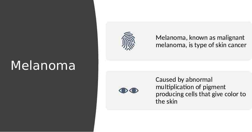
Melanoma, known as malignant melanoma, is type of skin cancer Melanoma Caused by abnormal multiplication of pigment producing cells that give color to the skin
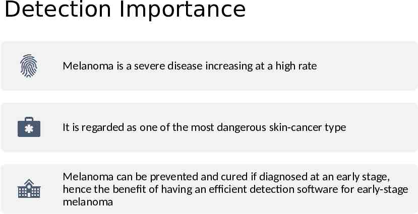
Detection Importance Melanoma is a severe disease increasing at a high rate It is regarded as one of the most dangerous skin-cancer type Melanoma can be prevented and cured if diagnosed at an early stage, hence the benefit of having an efficient detection software for early-stage melanoma
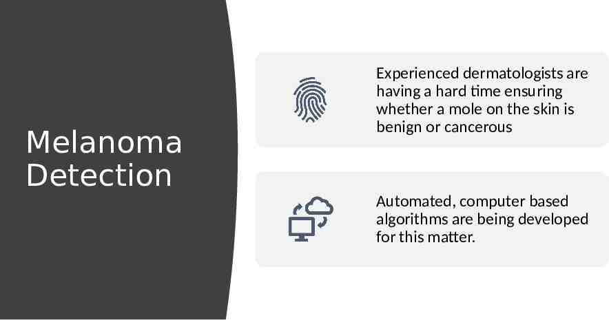
Melanoma Detection Experienced dermatologists are having a hard time ensuring whether a mole on the skin is benign or cancerous Automated, computer based algorithms are being developed for this matter.
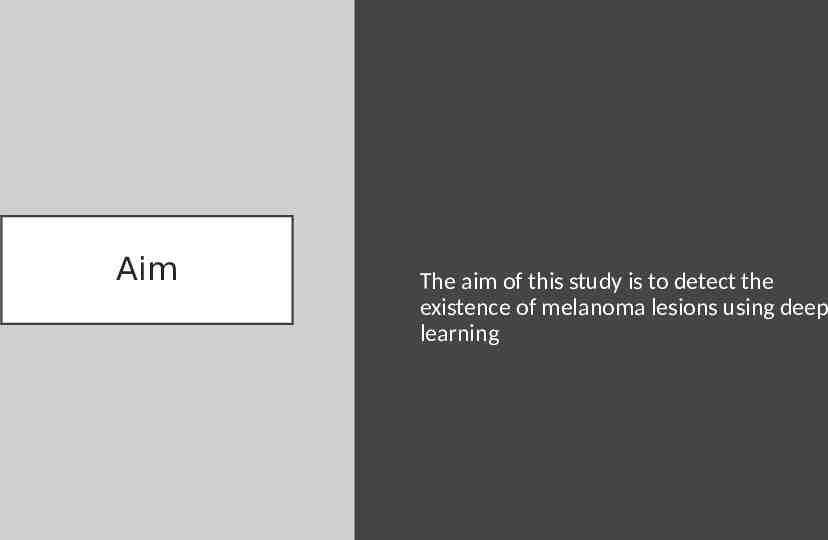
Aim The aim of this study is to detect the existence of melanoma lesions using deep learning
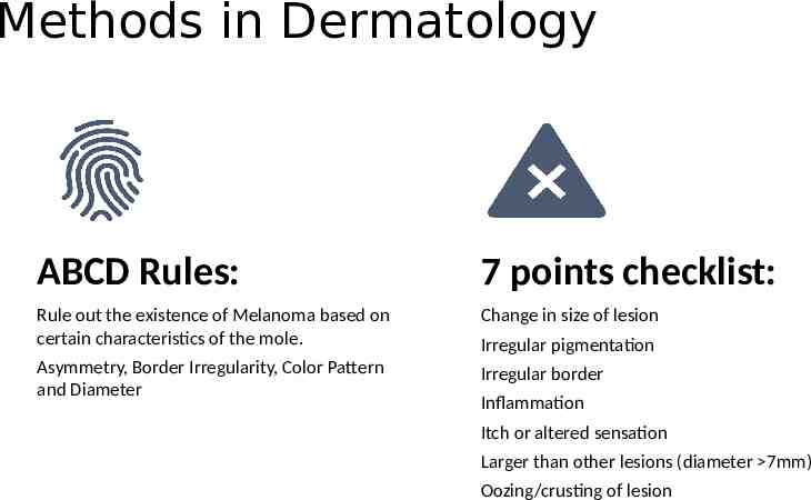
Methods in Dermatology ABCD Rules: 7 points checklist: Rule out the existence of Melanoma based on certain characteristics of the mole. Change in size of lesion Asymmetry, Border Irregularity, Color Pattern and Diameter Irregular border Irregular pigmentation Inflammation Itch or altered sensation Larger than other lesions (diameter 7mm) Oozing/crusting of lesion
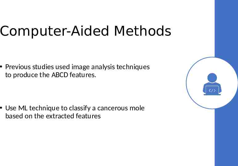
Computer-Aided Methods Previous studies used image analysis techniques to produce the ABCD features. Use ML technique to classify a cancerous mole based on the extracted features
Deep Learning Based Methods Deep learning has been used widely in different domains: Computer Vision Natural Language Processing Speech Recognition Etc. In this paper, the goal is to use deep learning for automated detection of skin cancer using digital images as input.
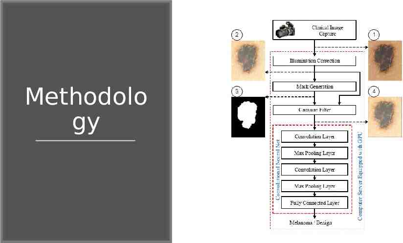
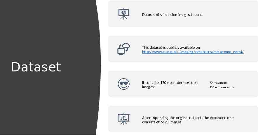
Image Acquisition http://www.cs.rug.nl/ imaging/databases/melanoma na evi/ Methodolo gy Image preprocessing for noise and illumination effects reduction Running Convolutional NN for classification
Methodolo gy
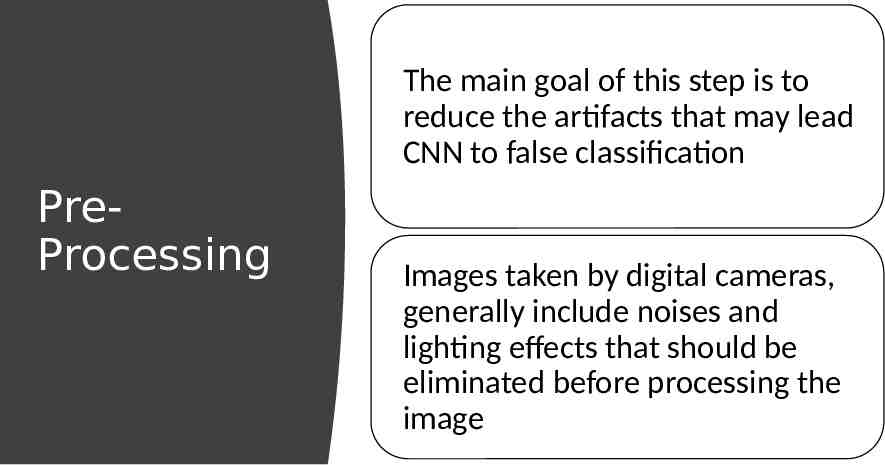
The main goal of this step is to reduce the artifacts that may lead CNN to false classification PreProcessing Images taken by digital cameras, generally include noises and lighting effects that should be eliminated before processing the image
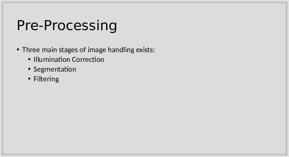
Pre-Processing Three main stages of image handling exists: Illumination Correction Segmentation Filtering
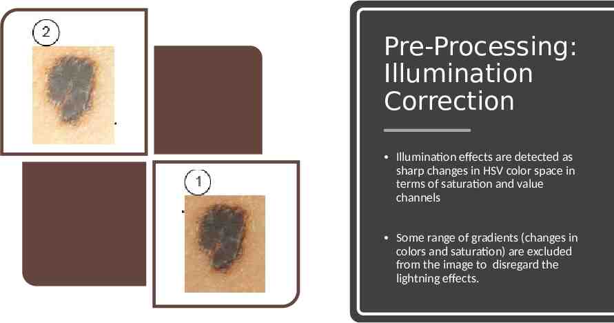
Pre-Processing: Illumination Correction Illumination effects are detected as sharp changes in HSV color space in terms of saturation and value channels Some range of gradients (changes in colors and saturation) are excluded from the image to disregard the lightning effects.
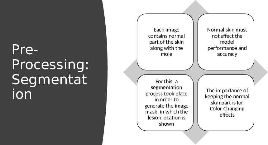
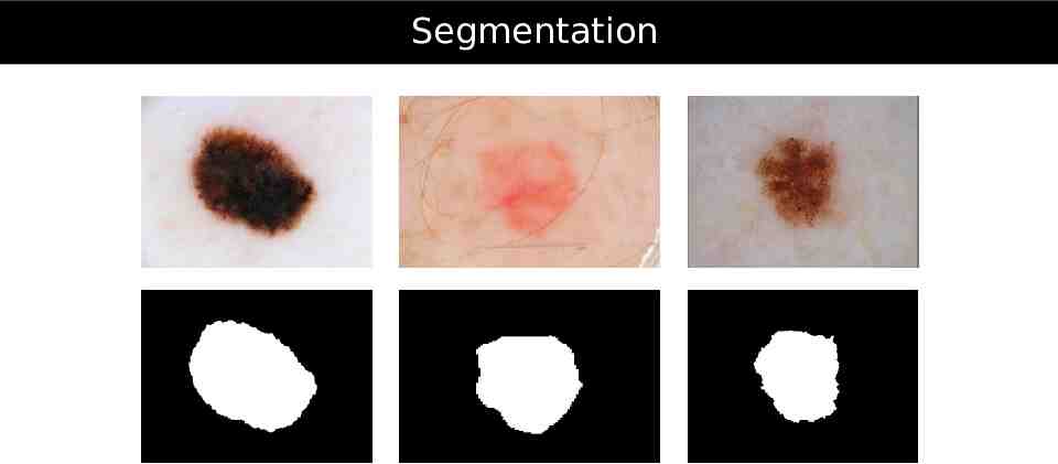
PreProcessing: Segmentat ion Each image contains normal part of the skin along with the mole Normal skin must not affect the model performance and accuracy For this, a segmentation process took place in order to generate the image mask, in which the lesion location is shown The importance of keeping the normal skin part is for Color Changing effects
Segmentation
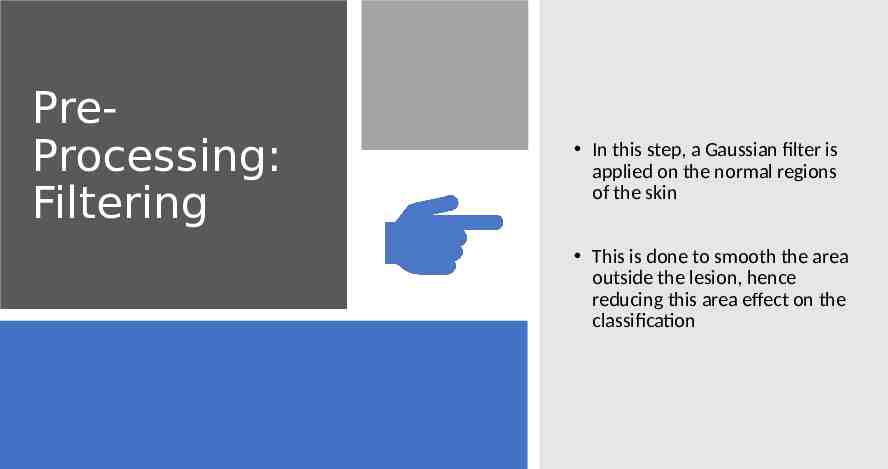
PreProcessing: Filtering In this step, a Gaussian filter is applied on the normal regions of the skin This is done to smooth the area outside the lesion, hence reducing this area effect on the classification
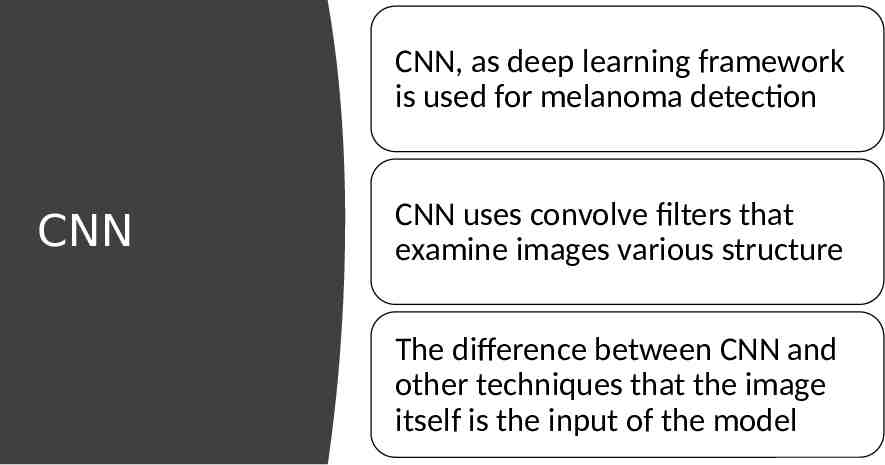
CNN, as deep learning framework is used for melanoma detection CNN CNN uses convolve filters that examine images various structure The difference between CNN and other techniques that the image itself is the input of the model
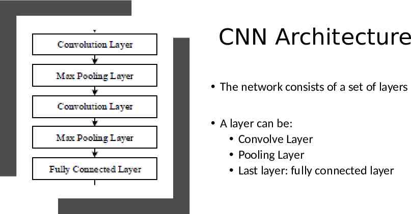
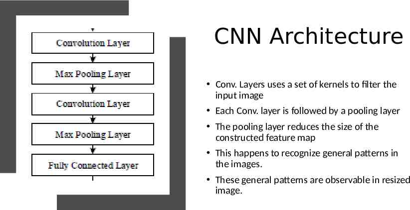
CNN Architecture The network consists of a set of layers A layer can be: Convolve Layer Pooling Layer Last layer: fully connected layer
CNN Architecture Conv. Layers uses a set of kernels to filter the input image Each Conv. layer is followed by a pooling layer The pooling layer reduces the size of the constructed feature map This happens to recognize general patterns in the images. These general patterns are observable in resized image.
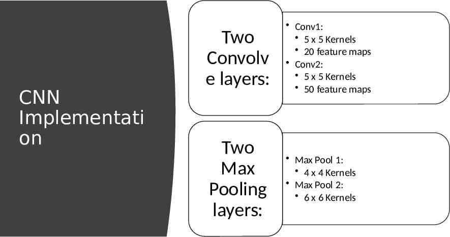
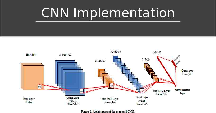
CNN Implementati on Two Convolv e layers: Two Max Pooling layers: Conv1: 5 x 5 Kernels 20 feature maps Conv2: 5 x 5 Kernels 50 feature maps Max Pool 1: 4 x 4 Kernels Max Pool 2: 6 x 6 Kernels
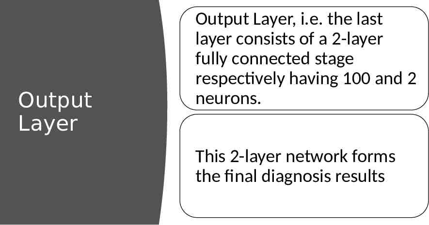
Output Layer Output Layer, i.e. the last layer consists of a 2-layer fully connected stage respectively having 100 and 2 neurons. This 2-layer network forms the final diagnosis results
CNN Implementation
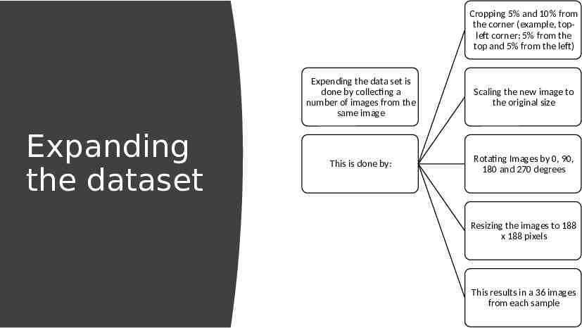
CNN needs a lot of training power in order to result in highly accurate predictive model Training CNN: Number of Sample issue Existing dataset consists of a low number of labeled images due to the fact that it is difficult to collect and label images This problem is fixed by expending the sample
Cropping 5% and 10% from the corner (example, topleft corner: 5% from the top and 5% from the left) Expanding the dataset Expending the data set is done by collecting a number of images from the same image Scaling the new image to the original size This is done by: Rotating Images by 0, 90, 180 and 270 degrees Resizing the images to 188 x 188 pixels This results in a 36 images from each sample
Dataset of skin lesion images is used. This dataset is publicly available on http://www.cs.rug.nl/ imaging/databases/melanoma naevi/ Dataset It contains 170 non - dermoscopic images: 70 melanoma 100 non-cancerous After expending the original dataset, the expanded one consists of 6120 images
Data is split into 80% for training and 20% for testing Experimen t Training set is fed to the network with batches Each batch size was 64, and the network was trained through 20000 iterations This is repeated 50 times to ensure stability, and the mean value of the results is reported
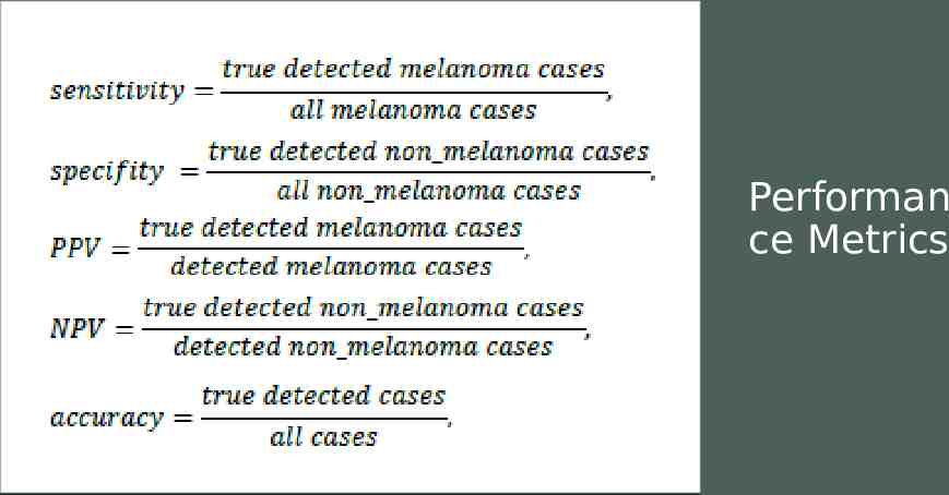
Performan ce Metrics
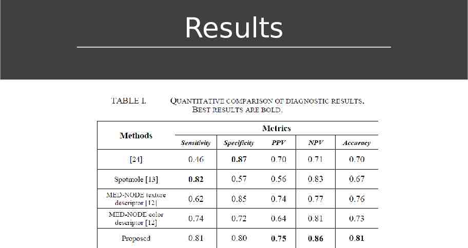
Comparing results Results were benchmarked against three previous studies done on the same data: (Zagrouba & Barhoumi, 2004) [24] is one of the first reported work, and used as baseline (Munteanu & Coocleam, 2009) [13] is an example of used commercial tools (Giotis et al, 2015) [12] classify cases in a semi-supervised framework
Results
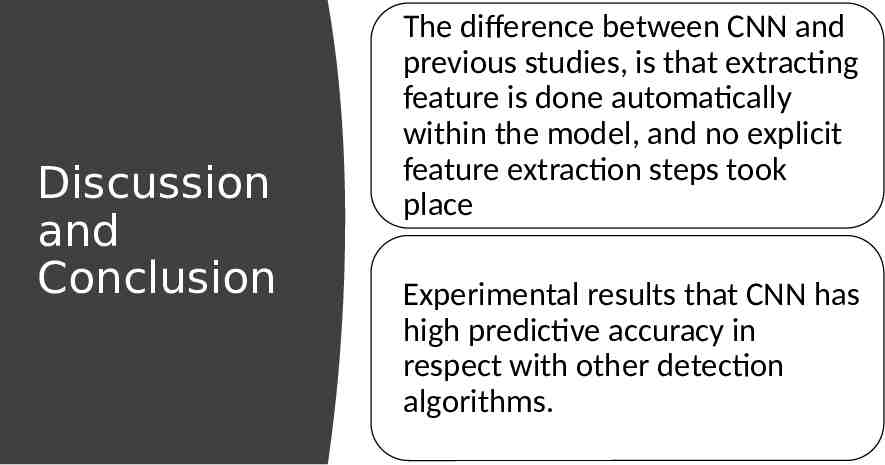
Discussion and Conclusion the used method in this paper (CNN) outperformed other state-of-the-art methods in terms of classification accuracy Deep Learned – based system is able to efficiently detect melanoma cases from non-cancerous ones Pre-processing of the images led to an increase of CNN accuracy
Discussion and Conclusion The difference between CNN and previous studies, is that extracting feature is done automatically within the model, and no explicit feature extraction steps took place Experimental results that CNN has high predictive accuracy in respect with other detection algorithms.
Thank You
Nasr-Esfahani, E., Samavi, S., Karimi, N., Soroushmehr, S. M. R., Jafari, M. H., Ward, K., & Najarian, K. (2016, August). Melanoma detection by analysis of clinical images using convolutional neural network. In 2016 38th Annual International Conference of the IEEE Engineering in Medicine and Biology Society (EMBC) (pp. 1373-1376). IEEE. Reference E. Zagrouba and W. Barhoumi, “A preliminary approach for the automated recognition of malignant melanoma, ” Image Analysis & Stereology, vol. 23, no. 2, pp. 121-135, 2004. C. Munteanu and S. Cooclea, “Spotmole – melanoma control system,” 2009. Available: http://www.spotmole.com/ I. Giotis, N. Molders, S. Land, M. Biehl, M. F. Jonkman and N. Petkov, “MEDNODE: A computer-assisted melanoma diagnosis system using nondermoscopic images,” Expert Systems with Applications, Elsevier, vol. 42, no. 19, pp. 6578-6585, 2015.