By Ojal Grover, Adorcille Abat, Patrick Trollope & Vijay Mahatma


62 Slides8.97 MB
By Ojal Grover, Adorcille Abat, Patrick Trollope & Vijay Mahatma
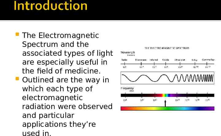
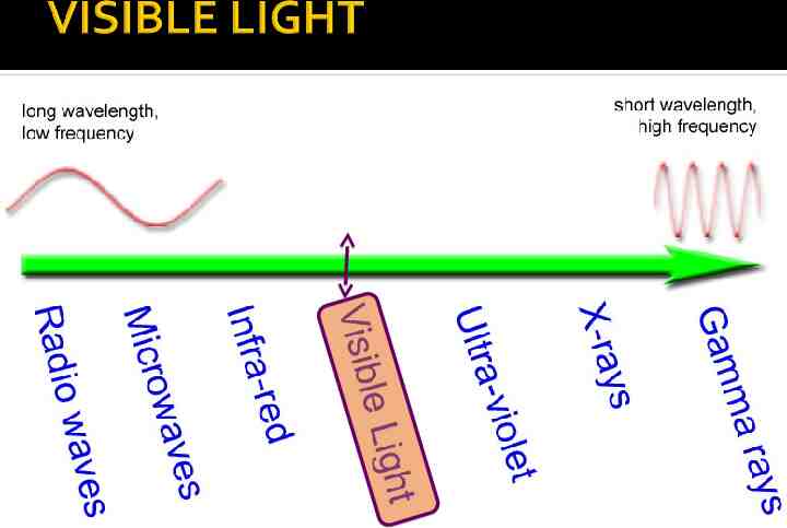
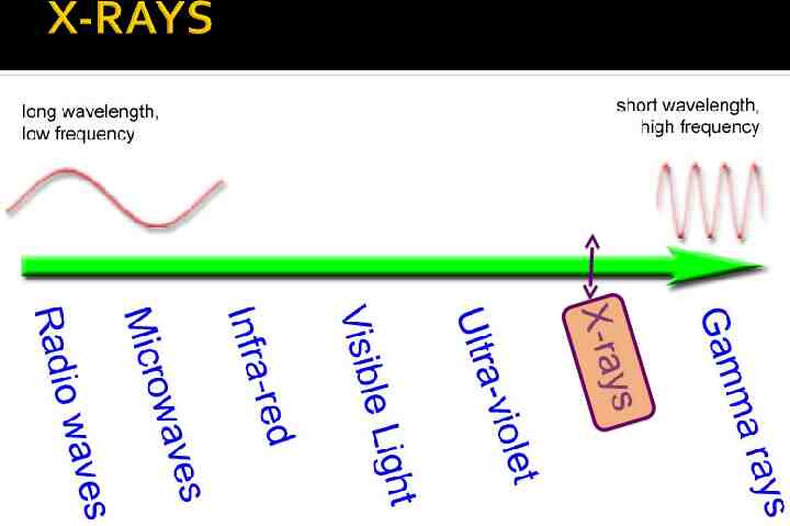
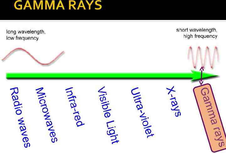
The Electromagnetic Spectrum and the associated types of light are especially useful in the field of medicine. Outlined are the way in which each type of electromagnetic radiation were observed and particular applications they’re used in.
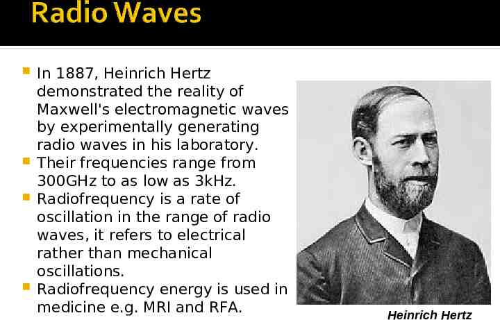
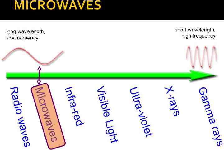
In 1887, Heinrich Hertz demonstrated the reality of Maxwell's electromagnetic waves by experimentally generating radio waves in his laboratory. Their frequencies range from 300GHz to as low as 3kHz. Radiofrequency is a rate of oscillation in the range of radio waves, it refers to electrical rather than mechanical oscillations. Radiofrequency energy is used in medicine e.g. MRI and RFA. Heinrich Hertz
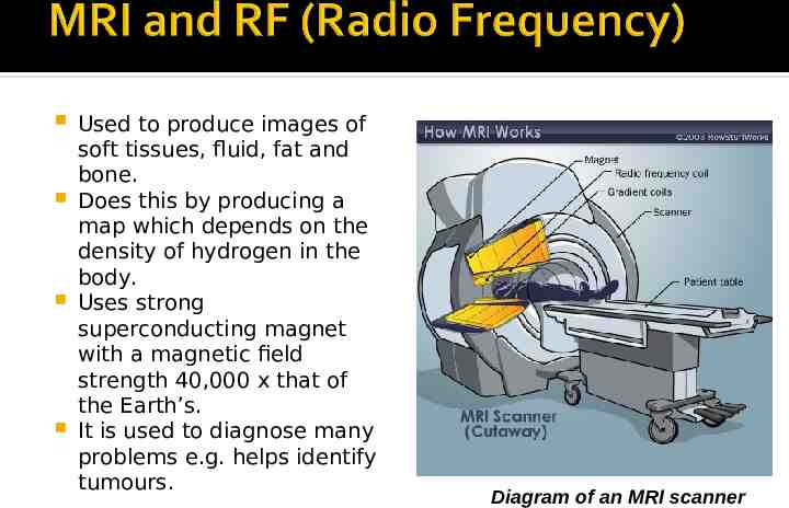
Used to produce images of soft tissues, fluid, fat and bone. Does this by producing a map which depends on the density of hydrogen in the body. Uses strong superconducting magnet with a magnetic field strength 40,000 x that of the Earth’s. It is used to diagnose many problems e.g. helps identify tumours. Diagram of an MRI scanner
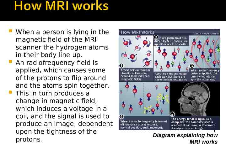
When a person is lying in the magnetic field of the MRI scanner the hydrogen atoms in their body line up. An radiofrequency field is applied, which causes some of the protons to flip around and the atoms spin together. This in turn produces a change in magnetic field, which induces a voltage in a coil, and the signal is used to produce an image, dependent upon the tightness of the protons. Diagram explaining how MRI works
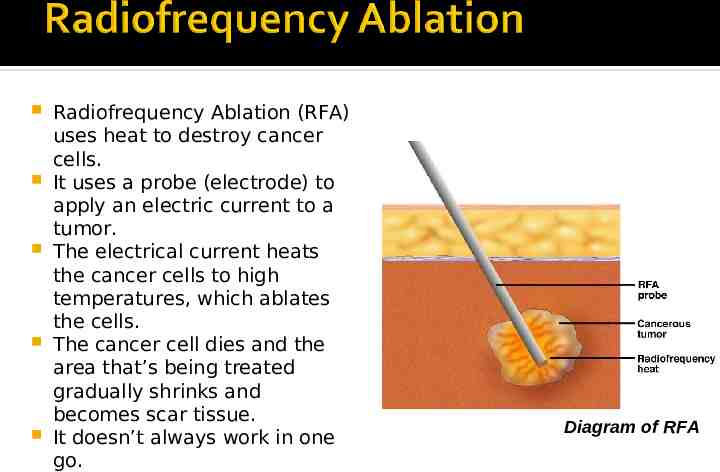
Radiofrequency Ablation (RFA) uses heat to destroy cancer cells. It uses a probe (electrode) to apply an electric current to a tumor. The electrical current heats the cancer cells to high temperatures, which ablates the cells. The cancer cell dies and the area that’s being treated gradually shrinks and becomes scar tissue. It doesn’t always work in one go. Diagram of RFA
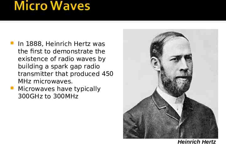
In 1888, Heinrich Hertz was the first to demonstrate the existence of radio waves by building a spark gap radio transmitter that produced 450 MHz microwaves. Microwaves have typically 300GHz to 300MHz Heinrich Hertz
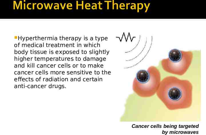
Hyperthermia therapy is a type of medical treatment in which body tissue is exposed to slightly higher temperatures to damage and kill cancer cells or to make cancer cells more sensitive to the effects of radiation and certain anti-cancer drugs. Cancer cells being targeted by microwaves
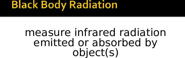
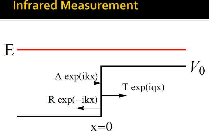
infrared radiation is emitted by all objects above absolute zero
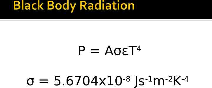
P AσεT4
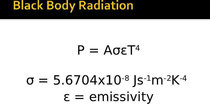
P AσεT4 σ 5.6704x10⁻⁸ Js-1m-2K-4
P AσεT4 σ 5.6704x10⁻⁸ Js-1m-2K-4 ε emissivity
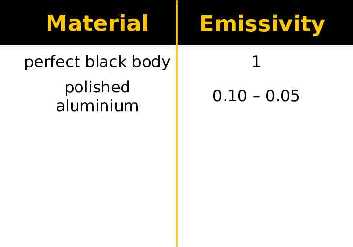
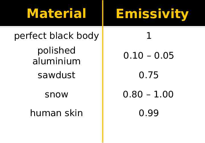
Material Emissivity
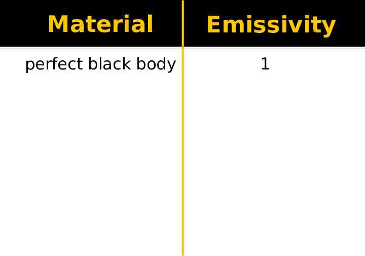
Material perfect black body Emissivity 1
Material Emissivity perfect black body 1 polished aluminium 0.10 – 0.05
Material Emissivity perfect black body 1 polished aluminium 0.10 – 0.05 sawdust 0.75 snow 0.80 – 1.00 human skin 0.99
measure infrared radiation emitted or absorbed by object(s)
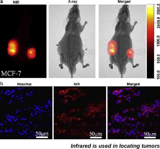
Infrared is used in locating tumors
1918 2009
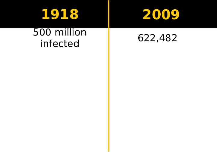
1918 500 million infected 2009 622,482
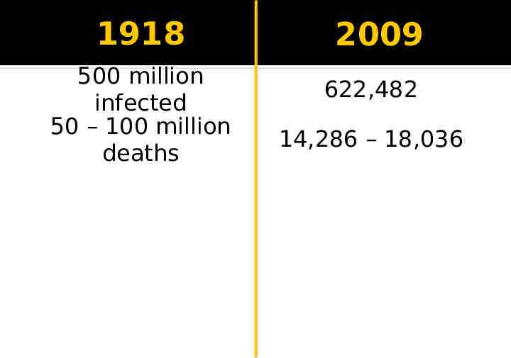
1918 500 million infected 50 – 100 million deaths 2009 622,482 14,286 – 18,036
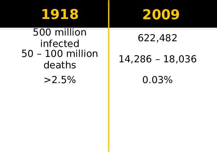
1918 500 million infected 50 – 100 million deaths 2.5% 2009 622,482 14,286 – 18,036 0.03%
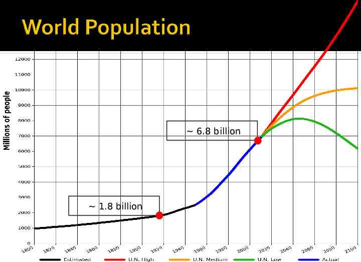
6.8 billion 1.8 billion
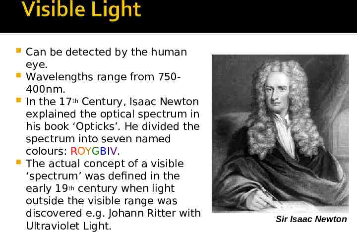
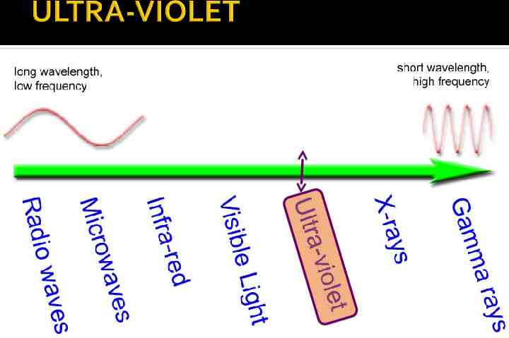
Can be detected by the human eye. Wavelengths range from 750400nm. In the 17th Century, Isaac Newton explained the optical spectrum in his book ‘Opticks’. He divided the spectrum into seven named colours: ROYGBIV. The actual concept of a visible ‘spectrum’ was defined in the early 19th century when light outside the visible range was discovered e.g. Johann Ritter with Ultraviolet Light. Sir Isaac Newton
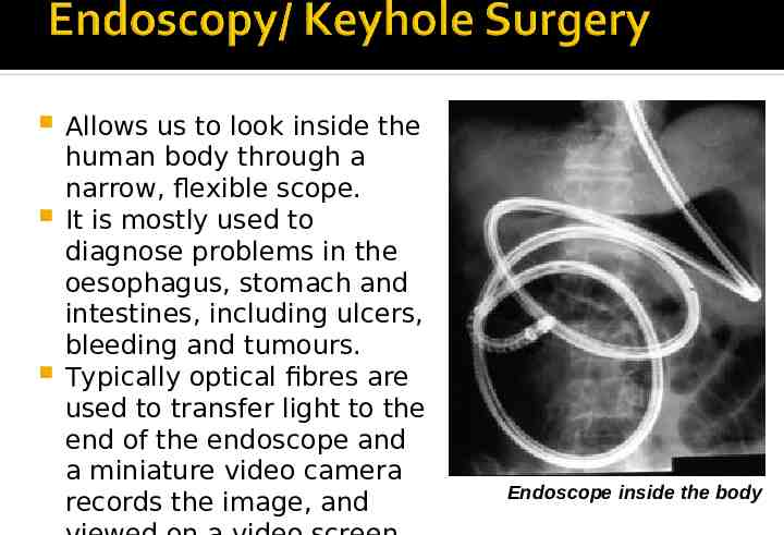
Allows us to look inside the human body through a narrow, flexible scope. It is mostly used to diagnose problems in the oesophagus, stomach and intestines, including ulcers, bleeding and tumours. Typically optical fibres are used to transfer light to the end of the endoscope and a miniature video camera records the image, and Endoscope inside the body
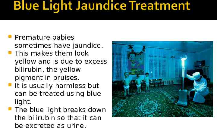
Premature babies sometimes have jaundice. This makes them look yellow and is due to excess bilirubin, the yellow pigment in bruises. It is usually harmless but can be treated using blue light. The blue light breaks down the bilirubin so that it can be excreted as urine.
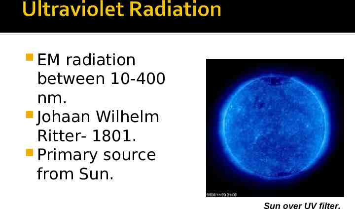
EM radiation between 10-400 nm. Johaan Wilhelm Ritter- 1801. Primary source from Sun. Sun over UV filter.
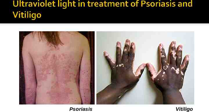
Psoriasis Vitiligo
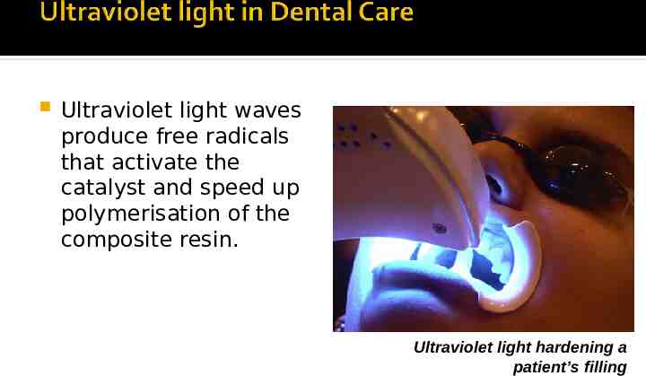
Ultraviolet light waves produce free radicals that activate the catalyst and speed up polymerisation of the composite resin. Ultraviolet light hardening a patient’s filling
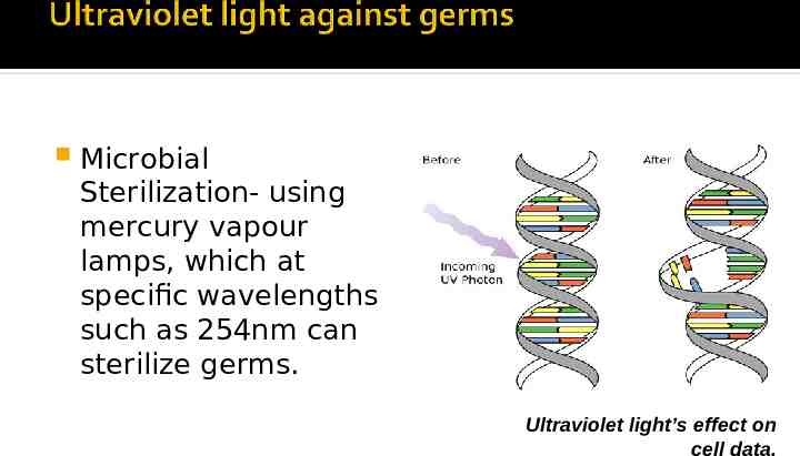
Microbial Sterilization- using mercury vapour lamps, which at specific wavelengths such as 254nm can sterilize germs. Ultraviolet light’s effect on cell data.
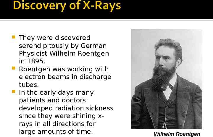
They were discovered serendipitously by German Physicist Wilhelm Roentgen in 1895. Roentgen was working with electron beams in discharge tubes. In the early days many patients and doctors developed radiation sickness since they were shining xrays in all directions for large amounts of time. Wilhelm Roentgen
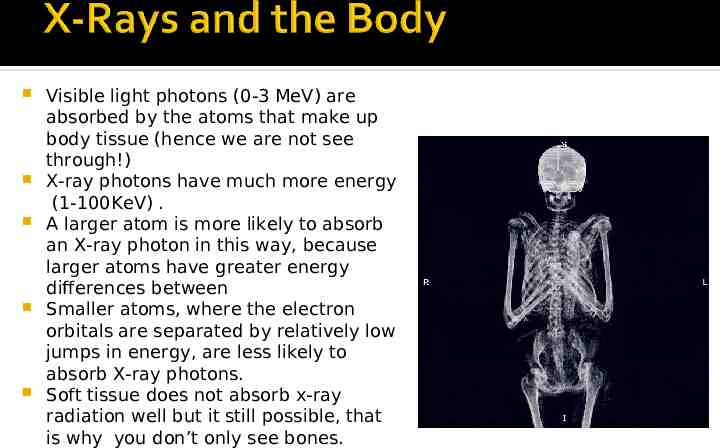
Visible light photons (0-3 MeV) are absorbed by the atoms that make up body tissue (hence we are not see through!) X-ray photons have much more energy (1-100KeV) . A larger atom is more likely to absorb an X-ray photon in this way, because larger atoms have greater energy differences between Smaller atoms, where the electron orbitals are separated by relatively low jumps in energy, are less likely to absorb X-ray photons. Soft tissue does not absorb x-ray radiation well but it still possible, that is why you don’t only see bones.
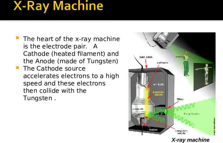
The heart of the x-ray machine is the electrode pair. A Cathode (heated filament) and the Anode (made of Tungsten) The Cathode source accelerates electrons to a high speed and these electrons then collide with the Tungsten . X-ray machine
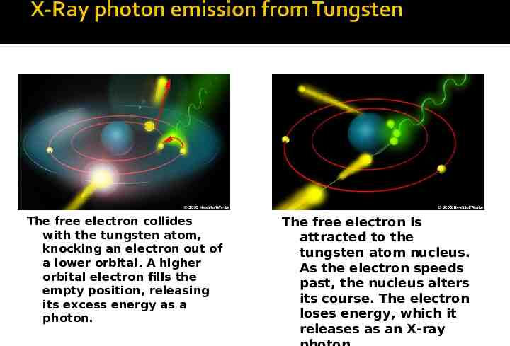
The free electron collides with the tungsten atom, knocking an electron out of a lower orbital. A higher orbital electron fills the empty position, releasing its excess energy as a photon. The free electron is attracted to the tungsten atom nucleus. As the electron speeds past, the nucleus alters its course. The electron loses energy, which it releases as an X-ray
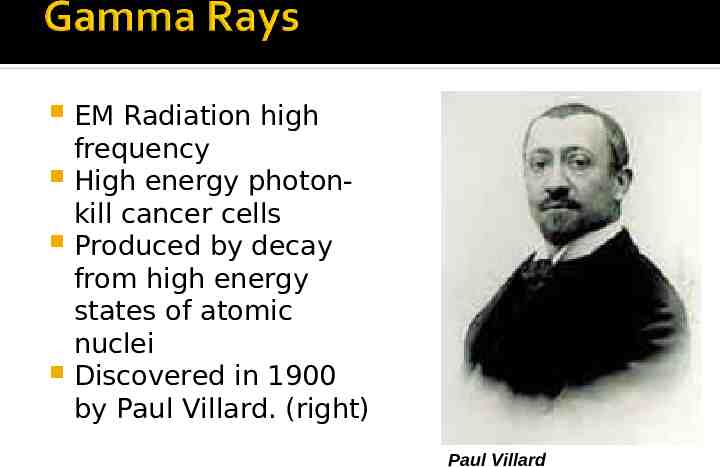
EM Radiation high frequency High energy photonkill cancer cells Produced by decay from high energy states of atomic nuclei Discovered in 1900 by Paul Villard. (right) Paul Villard
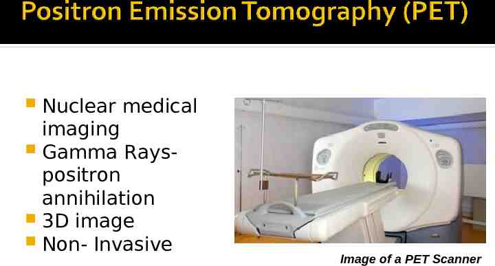
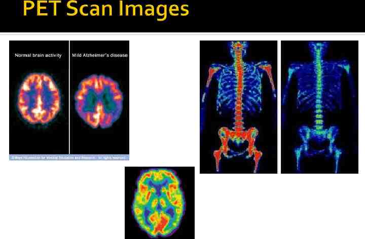
Nuclear medical imaging Gamma Rayspositron annihilation 3D image Non- Invasive Image of a PET Scanner
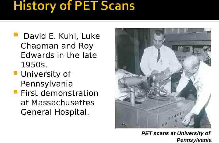
David E. Kuhl, Luke Chapman and Roy Edwards in the late 1950s. University of Pennsylvania First demonstration at Massachusettes General Hospital. PET scans at University of Pennsylvania
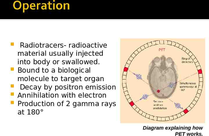
Radiotracers- radioactive material usually injected into body or swallowed. Bound to a biological molecule to target organ Decay by positron emission Annihilation with electron Production of 2 gamma rays at 180 Diagram explaining how PET works.
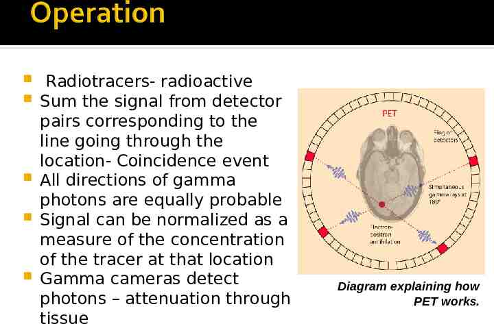
Radiotracers- radioactive Sum the signal from detector pairs corresponding to the line going through the location- Coincidence event All directions of gamma photons are equally probable Signal can be normalized as a measure of the concentration of the tracer at that location Gamma cameras detect photons – attenuation through tissue Diagram explaining how PET works.
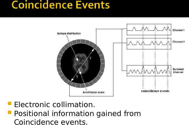
Electronic collimation. Positional information gained from Coincidence events.
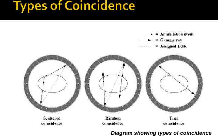
Diagram showing types of coincidence
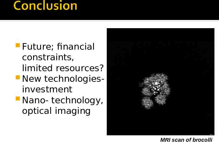
Future; financial constraints, limited resources? New technologiesinvestment Nano- technology, optical imaging MRI scan of brocolli
By Ojal Grover, Adorcille Abat, Patrick Trollope & Vijay Mahatma