Assessment and Management of Patients with Endocrine Disorders By
78 Slides496.00 KB
Assessment and Management of Patients with Endocrine Disorders By Linda Self
Glands of the Endocrine System Hypothalamus Posterior Pituitary Anterior Pituitary Thyroid Parathyroids Adrenals Pancreatic islets Ovaries and testes
Hypothalamus Releasing and inhibiting hormones Corticotropin-releasing hormone Thyrotropin-releasing hormone Growth hormone-releasing hormone Gonadotropin-releasing hormone Somatostatin- -inhibits GH and TSH
Anterior Pituitary Growth Hormone- Adrenocorticotropic hormone Thyroid stimulating hormone Follicle stimulating hormone—ovary in female, sperm in males Luteinizing hormone—corpus luteum in females, secretion of testosterone in males Prolactin—prepares female breasts for lactation
Posterior Pituitary Antidiuretic Hormone Oxytocin—contraction of uterus, milk ejection from breasts
Adrenal Cortex Mineralocorticoid—aldosterone. Affects sodium absorption, loss of potassium by kidney Glucocorticoids—cortisol. Affects metabolism, regulates blood sugar levels, affects growth, antiinflammatory action, decreases effects of stress Adrenal androgens—dehydroepiandrosterone and androstenedione. Converted to testosterone in the periphery.
Adrenal Medulla Epinephrine and norepinephrine serve as neurotransmitters for sympathetic system
Thyroid Follicular cells—excretion of triiodothyronine (T3) and thyroxine (T4)—Increase BMR, increase bone and calcium turnover, increase response to catecholamines, need for fetal G&D Thyroid C cells—calcitonin. Lowers blood calcium and phosphate levels
Parathyroid Parathyroid hormone—regulates serum calcium
Pancreatic Islet cells Insulin Glucagon—stimulates glycogenolysis and glyconeogenesis Somatostatin—decreases intestinal absorption of glucose
Kidney 1, 25 dihydroxyvitamin D—stimulates calcium absorption from the intestine Renin—activates the RAAS Erythropoietin—Increases red blood cell production
Ovaries Estrogen Progesterone—inportant in menstrual cycle,*maintains pregnancy,
Testes Androgens, testosterone—secondary sexual characteristics, sperm production
Thymus Releases thymosin and thymopoietin Affects maturation of T lymphocetes
Pineal Melatonin Affects sleep, fertility and aging
Prostaglandins Work locally Released by plasma cells Affect fertility, blood clotting, body temperature
Assessment Health history—energy level, hand and foot size changes, headaches, urinary changes, heat and cold intolerance, changes in sexual characteristics, personality changes, others Physical assessment—appearance including hair distribution, fat distribution, quality of skin, appearance of eyes, size of feet and hands, peripheral edema, facial puffiness, vital signs
Diagnostic Evaluation Serum levels of hormones Detection of antibodies against certain hormones Urinary tests to measure by-products (norepinephrine, metanephrines, dopamine) Stimulation tests—determine how an endocrine gland responds to stimulating hormone. If the hormone responds, then the problem lies w/hypothalmus or pituitary Suppression tests—tests negative feedback systems that control secretion of hormones from the hypothalamus or pituitary.
Disorders of the Pituitary Pituitary Tumors Eosinophilic tumors may result in gigantism or in acromegaly. May suffer from severe headaches, visual disturbances, decalcification of the bone, endocrine disturbances Basophilic tumors may cause Cushing’s syndrome w/features of hyperadrenalism, truncal obesity, amenorrhea, osteoporosis Chromophobic tumors—90% of pituitary tumors. Present with lowered BMR, obesity, somnolence, scant hair, low body temp, headaches, visual changes
Growth hormone deficiency in childhood will result in primary dwarfism.
Pituitary Tumors—Assessment and Diagnostic Findings H&P Vision tests CT, MRI Serum levels of pituitary hormones, others
Diabetes Insipidus Deficiency of ADH Excessive thirst, large volumes of dilute urine Can occur secondary to brain tumors, head trauma, infections of the CNS, and surgical ablation or radiation Nephrogenic DI—relates to failure of the renal tubules to respond to ADH. Can be related to hypokalemia, hypercalcemia and to medications (lithium demeocycline)
Manifestations Excessive thirst Urinary sp. gr. of 1.001.1.005
Assessment and Diagnostic Findings Fluid deprivation test—withhold fluids for 8-12 hours. Weigh patient frequently. Inability to slow down the urinary output and fail to concentrate urine are diagnostic. Stop test if patient is tachycardic or hypotensive Trial of desmopressin and IV hypertonic saline Monitor serum and urine osmolality and ADH levels
Pharmacologic Tx and Nursing Management DDAVP—intranasal bid Can be given IM if necessary. Every 24-96h. Can cause lipodystrophy. Can also use Diabenese and thiazide diuretics in mild disease as they potentiate the action of ADH If renal in origin—thiazide diuretics, NSAIDs (prostaglandin inhibition) and salt depletion may help Educate patient about actions of medications, how to administer meds, wear medic alert bracelet
SIADH Excessive ADH secretion Retain fluids and develop a dilutional hyponatremia Often non-endocrine in origin—such as bronchogenic carcinoma Causes: Disorders of the CNS like head injury, brain surgery, tumors, infections or medications like vincristine, phenothiazines, TCAs or thiazide diuretics Meds can either affect the pituitary or increase sensitivity to renal tubules to ADH Management: eliminate cause, give diuretics (Lasix), fluid restriction, I&O, daily wt., lab chemistries
SIADH Restoration of electrolytes must be gradual May use 3% NaCl in conjunction with Lasix
Thyroid T3 and T4 Need iodine for synthesis of hormones—excess will result in adaptive decline in utilization called the WolfChaikoff mechanism Thyroid is controlled by TSH Cellular metabolism, brain development, normal growth, affect every organ in the body T3 is five times as potent as T4 Calcitonin—secreted in response to high levels of serum calcium, increases deposition in the bone
Thyroid Inspect gland Observe for goiter Check TSH, serum T3 and T4 T3 resin uptake test useful in evaluating thyroid hormone levels in patients who have received diagnostic or therapeutic dose of iodine. Estrogens, Dilantin, Tagamet, Heparin, amiodarone, PTU,steroids and Lithium can cloud the accuracy T3 more accurate indicator of hyperthyroidism according to text
Thyroid Antibodies seen in Hashimoto’s, Grave’s and other auto-immune problems. Radioactive iodine uptake test measures rate of iodine uptake. Patients with hyperthyroidism exhibit a high uptake, hypothyroidism will have low uptake Thyroid scan—helps determine the location, size, shape and size of gland. “Hot” areas (increased function) and “cold” areas (decreased function) can assist in diagnosis.
Nursing Implications Be aware of meds patient is taking (see list in text) that can affect accuracy of testing Also be aware if patient is taking multivitamins and food supplements
Hypothyroidism Most common cause is Hashimoto’s thyroiditis Common in those previously treated for hyperthyroidism Atrophy of gland with aging Medications like lithium, iodine compounds, antithyroid meds can cause Radiation treatments to head and neck Infiltrative diseases like amyloidosis, scleroderma Iodine deficiency and excess Hypothalamic or pituitary abnormality More common in women, especially over age 50
Manifestations From mild symptoms to myxedema Myxedema –accumulation of mucopolysaccharides in sc and interstitial tissues. Is the extreme form of hypothyroidism. Can progress to shock. S/S—fatigue, hair loss, dry skin, brittle nails, numbness and tingling of the fingers, amenorrhea, weight gain, decreased heart rate and temperature, lassitude, cognitive changes, elevated cholesterol levels, constipation, hypotension
Pharmacologic Management of hypothyroidism Levothyroxine is preferred agent Dosage is based on TSH Desiccated thyroid used infrequently due to inconsistent dosing Angina can occur when thyroid replacement is initiated as it enhances effects of cardiovascular catecholamines (in pt. w/pre-existent CAD). Start at low dose. Hypnotics and sedatives may have profound effects on sensorium
Management in Myxedema Cautious fluid replacement Glucose to restore to normal glycemic levels Avoid rapid overheating due to increased oxygen demands but keep warm May give levothyroxine intravenously With recovery, Modify activity High fiber foods Home health for follow-up
Hyperthyroidism Extreme form is Grave’s disease Caused by thyroiditis, excessive amount thyroid hormone, abnormal output by immunoglobulins Is more common in women
Manifestations of hyperthyroidism Thyrotoxicosis—nervousness, irritable, apprehensive, palpitations, heat intolerance, skin flushing, tremors, possibly exophthalmos Have an increased sensitivity to catecholamines Can occur after irradiation or presence of a tumor
Assessment and Diagnosis Thyroid thrill and or bruit may be present Thyroid may be enlarged Decreased TSH, increased free T4 and an increased radioactive iodine uptake
Management Reduce thyroid hyperactivity—usually use radioactive iodine, antithyroid meds or surgery) Beta blockers Can be relapse with antithyroid meds
Pharmacologic Therapy Irradiation with administration of radioisotope iodine 131—initially may cause an acute release of thyroid hormones. Should monitor for thyroid storm S/S of thyroid storm—high fever. Tachycardia, delirium, chest pain, dyspnea, palpitations, weight loss, diarrhea, abdominal pain Management of thyroid storm—oxygen, IV fluids with dextrose, hypothermic measures, steroids to treat shock or adrenal deficiency, iodine to decrease output of T4, beta blockers, PTU or Tapazole impedes formation of thyroid hormone and blocks conversion of T4 to T3
Antithyroid Medications PTU—propylthiouracil—blocks synthesis of hormones Tapazole (methimazole)—blocks synthesis of hormones. More toxic than PTU. Sodium Iodide-suppresses release of thyroid hormone SSKI (saturated solution of potassium chloride)– suppresses release of hormones and decreases vascularity of thyroid. Can stain teeth Dexamethazone—suppresses release of thyroid hormones
Surgical Management Reserved for special circumstances, e.g. large goiters, those who cannot take antithyroid meds, or who need rapid normalization Subtotal thyroidectomy Before surgery, give PTU until s/s of hyperthyroidism have disappeared Iodine may be used to decrease vascularity
Nursing Management Reassurance r/t the emotional reactions experienced May need eye care if has exophthalmos Maintain normal body temperature Adequate caloric intake Managing potential complications such as dysrhythmias and tachycardias Educate about potential s/s of hypothyroidism following any antithyroid tx.
Parathyroid Glands Parathormone maintains sufficient serum calcium levels Excess calcium can bind with phosphate and precipitate in various organs, can cause pancreatitis Hyperparathyroidism will cause bone decalcification and development of renal calculi More common in women Secondary hyperparathyroidism occurs in those with chronic renal failure and renal rickets secondary to excess phosphorus retention (and increased parathormone secretion)
Manifestations of Hyperparathyroidism May be asymptomatic Apathy, fatigue, muscle weakness, nausea, vomiting, constipation, hypertension and cardiac dysrhythmias Excess calcium in the brain can lead to psychoses Renal lithiasis can lead to renal damage and even failure Demineralization of bones with back and joint pain, pain on weight bearing, pathologic fractures Peptic ulcers and pancreatitis can also occur
Assessment and Diagnostic Findings Persistent elevated calcium levels Elevated serum parathormone level Bone studies will reveal decreased density Double antibody parathyroid hormone test is used to distinguish between primary hyperparathyroidism and malignancy Ultrasound, MRI, thallium scan, fine needle biopsy also can be used to localize cysts, adenomas, or hyperplasia
Management Recommended treatment for hyperparathyroidism is surgical removal Hydration therapy necessary to prevent renal calculi Avoid thiazide diuretics as they decrease renal excretion of calcium Increase mobility to promote bone retention of calcium Avoid restricted or excess calcium in the diet Fluids, prune juice and stool softeners to prevent constipation Watch for s/s of tetany postsurgically (numbness, tingling, carpopedal spasms) as well as cardiac dysrhythmias and hypotension
Hypercalcemic crisis Seen with levels greater than 15mg/dL Can result in life-threatening neurologic, cardiovascular and renal symptoms Treatments include: hydration, loop diuretics to promote excretion of calcium, phosphate therapy to promote calcium deposition in bone and reducing GI absorption of calcium Give calcitonin or mithramycin to decrease serum calcium levels quickly
Hypoparathyroidism Seen most often following removal of thyroid gland, parathyroid glands or following radical neck surgery Deficiency of parathormone results in increased bone phosphate and decreased blood calcium levels In absence of parathormone, there is decreased intestinal absorption of dietary calcium and decreased resorption of calcium from bone and through kidney tubules
Clinical Manifestations of Hypoparathyroidism Irritability of neuromuscular system Tetany—hypertonic muscle contractions , numbnes, tingling, cramps in extremities, laryngeal spasm, bronchospasm, carpopedal spasm ( flexion of the elbows and wrists, dorsiflexion of the feet), seizures
Assessment and Diagnostic Findings Trousseau’s sign—can check with a BP cuff Chvostek’s sign—tapping over facial nerve causes spasm of the mouth, nose and eye Lab studies may reveal calcium levels of 5-6 mg/dL or lower Serum phosphate levels will be decreased
Management of Hypoparathyroidism Restore calcium level to 9-10 mg/dL May need to give IV calcium gluconate for immediate treatment Use of parathormone IV reserved for extreme situations due to the probability of allergic reactions Monitor calcium levels May need bronchodilators and even ventilator assistance Diet high in calcium and low in phosphorus; thus, avoid milk products, egg yolk and spinach.
Management of Hypoparathyroidism Keep calcium gluconate at bedside Ensure has IV access Cardiac monitoring Care of postoperative patients who have undergone thyroid surgery, parathyroidectomy or radical neck surgery. Be watchful for signs of tetany, seizures, and respiratory difficulties
Adrenals--Pheochromocytoma Usually benign tumor Originates from the chromaffin cells of the adrenal medulla Any age but usu. Between 40-50 years old Can be familial 10% are malignant May be associated with thyroid carcinoma or parathyroid hyperplasia or tumor
Clinical Manifestations Headache, diaphoresis, palpitations, hypertension May have hyperglycemia related to excess epinephrine secretion Tremors, flushing and anxiety as well Blurring of vision Feeling of impending doom BPs exceeding 250/150 have occurred
Assessment and Diagnostic Findings Associated with the 5 H’s—hypertension, headache, hyperhidrosis, hypermetabolism and hyperglycemia Urinary catecholamines and metanephrine are direct and conclusive tests Serum epinephrine and norepinephrine levels will be elevated Urinary vanillymandelic acid also diagnostic Must avoid coffee, tea, bananas, chocolate, vanilla and ASA, nicotine, amphetamines, decongestants before 24h urine testing Clonidine suppression test—in normal individual, would block catecholamine release Imaging studies
Management Bedrest Elevated HOB ICU Nipride Calcium channel blockers and Beta blockers Surgical management (manipulation of the tumor can cause excessive release of catecholamines) Steroid therapy if adrenalectomy performed Hypotension and hypoglycemia can occur post-op
Addison’s Disease Adrenocortical insufficiency Autoimmune or idiopathic atrophy Can be caused by inadequate ACTH from pituitary Therapeutic use of steroids
Manifestations Muscle weakness Anorexia Dark pigmentation Hypotension Hypoglycemia Low sodium levels High potassium levels Can result in Addisonian crisis
Addisonian crisis Circulatory shock Pallor, apprehension, weak&rapid pulse, rapid respirations and low blood pressure Headache, nausea, abdominal pain and diarrhea Can be brought on by overexertion, exposure to cold, acute infection, decrease in salt intake
Assessment and Diagnostic Findings Early morning serum cortisol and plasma ACTH are performed. Will distinguish between primary and secondary adrenal insufficiency. In primary, will have elevated ACTH levels and below normal cortisol levels. If the adrenal cortex is not stimulated by the pituitary, a normal response to doses of exogenous ACTH (see text) Blood sugar levels and electrolyte values
Management Restore circulatory status—fluids, steroids May need antibiotics if infection precipitated crisis May need lifelong steroid therapy and mineralocorticoid therapy May need additional salt intake Check orthostatics Daily weights Aware that stressors can precipitate crises Medic alert bracelet or similar identification of history
Cushing’s Syndrome Results from excessive adrenocortical activity May be related to excessive use of corticosteroid medications or due to hyperplasia of the adrenal cortex Oversecretion of corticosteroids can also be caused by pituitary tumor Can be caused by bronchogenic carcinoma or other malignancy
Manifestations of Cushing’s syndrome Cataracts, glaucoma Hypertension, heart failure Truncal obesity, moon face, buffalo hump, sodium retention, hypokalemia, hyperglycemia, negative nitrogen balance, altered calcium metabolism Decreased inflammatory responses, impaired wound healing, increased susceptibility to infections Osteoporosis, compression fractures Peptic ulcers, pancreatitis Thinning of skin, striae, acne Mood alterations
Assessment and Diagnostic Findings Overnight dexamethasone suppression test frequently used for diagnosis Administered at 11pm and cortisol level checked at 8am Suppression of cortisol to less than 5mg/dL indicates normal functioning Measurement of plasma ACTH (radioimmunoassay) in conjunction with dexamethasone suppression test helps distinguish pituitary vs. ectopic sites of ACTH. MRI, CT and CT also help detect tumors of adrenal or pituitary
Medical Management If pituitary source, may warrant transphenoidal hypophysectomy Radiation of pituitary also appropriate Adrenalectomy may be needed in case of adrenal hypertrophy Temporary replacement therapy with hydrocortisone or Florinef Adrenal enzyme reducers may be indicated if source if ectopic and inoperable. Examples include: ketoconazole, mitotane and metyrapone. If cause is r/t excessive steroid therapy, tapering slowly to a minimum dosage may be appropriate.
Primary Aldosteronism or Conn’s Syndrome Excessive aldosterone secondary to adrenal tumor retain sodium and excrete potassium Results in alkalosis Hypertension—universal sign of hyperaldosteronism Inability of kidneys to concentrate the urine Serum becomes concentrated Excessive thirst Hypokalemia interferes with insulin secretion thus will have glucose intolerance as well
Assessment and Diagnostic Findings High sodium Low potassium level High serum aldosterone level Low renin level Aldosterone excretion rate after salt loading is diagnostic for primary aldosteronism Renin-aldosterone stimulation test
Management Surgical removal of tumor Correct hypokalemia Usual postoperative care with abdominal surgery Administer steroids Fluids Monitoring of blood sugar Control of hypertension with spironolactone
Corticosteroid Therapy Hydrocortisone--Cortisol Cortisone--Cortate Prednisone--Deltasone Prednisolone-Prelone Triamcinolone--Kenalog Betamethasone--Celestone Fludrocortisone (contains both mineralocorticoid and glucocorticoid) Florinef
Indications RA Asthma MS COPD exacerbations Lupus Other autoimmune disorders Dermatologic disorders
Dosing Lowest dose Limited duration Best time to give dose is in early morning between 7-8 am Need to taper off med to allow normal return of renal function
Side Effects of Steroids Hypertension, thrombophlebitis, accelerated atherosclerosis Increased risk of infection Glaucoma and corneal lesions Muscle wasting, poor wound healing, osteoporosis, pathologic fractures Hyperglycemia, steroid withdrawal syndrome Moon face, weight gain, acne
Case Study 1 35 year old male presents with BP of 188/112 at a yearly physical exam. Previous exams noted blood pressures of 160/94 and 158/92. On questioning, patient admits to twice a month episodes of apprehension, severe headache, perspiration, rapid heartbeat, and facial pallor. These episodes had an abrupt onset and lasted 10-15 minutes. Routine hematology and chemistry studies are wnl and chest xray and ECG are normal. What is your impression? What labs would you draw?
Case Study 2 50 year old woman presents with enlargement of left anterior neck. She has noted increased appetite over the past month with no weight gain, and more frequent bowel movements over the same period. Patient feels jittery at times, experiences palpitations and feels “hot” a lot recently. She is 5’8” tall and weighs 150#. Heart rate is 110 and blood pressure is 110/76. What might be this patient’s problem? What lab tests might you draw?
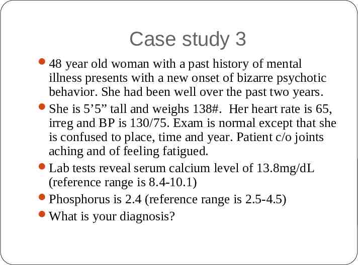
Case study 3 48 year old woman with a past history of mental illness presents with a new onset of bizarre psychotic behavior. She had been well over the past two years. She is 5’5” tall and weighs 138#. Her heart rate is 65, irreg and BP is 130/75. Exam is normal except that she is confused to place, time and year. Patient c/o joints aching and of feeling fatigued. Lab tests reveal serum calcium level of 13.8mg/dL (reference range is 8.4-10.1) Phosphorus is 2.4 (reference range is 2.5-4.5) What is your diagnosis?
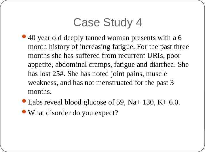
Case Study 4 40 year old deeply tanned woman presents with a 6 month history of increasing fatigue. For the past three months she has suffered from recurrent URIs, poor appetite, abdominal cramps, fatigue and diarrhea. She has lost 25#. She has noted joint pains, muscle weakness, and has not menstruated for the past 3 months. Labs reveal blood glucose of 59, Na 130, K 6.0. What disorder do you expect?
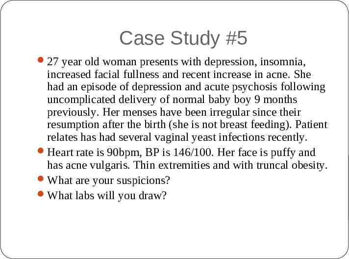
Case Study #5 27 year old woman presents with depression, insomnia, increased facial fullness and recent increase in acne. She had an episode of depression and acute psychosis following uncomplicated delivery of normal baby boy 9 months previously. Her menses have been irregular since their resumption after the birth (she is not breast feeding). Patient relates has had several vaginal yeast infections recently. Heart rate is 90bpm, BP is 146/100. Her face is puffy and has acne vulgaris. Thin extremities and with truncal obesity. What are your suspicions? What labs will you draw?