Circulatory unit 8 ppt#3 Major Arteries and Veins Blood pressure


30 Slides3.73 MB
Circulatory unit 8 ppt#3 Major Arteries and Veins Blood pressure
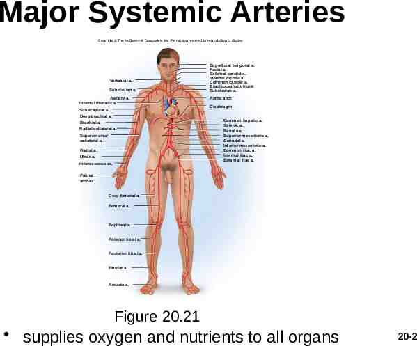
Major Systemic Arteries Copyright The McGraw-Hill Companies, Inc. Permission required for reproduction or display. Vertebral a. Subclavian a. Axillary a. Internal thoracic a. Subscapular a. Deep brachial a. Brachial a. Radial collateral a. Superior ulnar collateral a. Radial a. Ulnar a. Interosseous aa. Superficial temporal a. Facial a. External carotid a. Internal carotid a. Common carotid a. Brachiocephalic trunk Subclavian a. Aortic arch Diaphragm Common hepatic a. Splenic a. Renal aa. Superior mesenteric a. Gonadal a. Inferior mesenteric a. Common iliac a. Internal iliac a. External iliac a. Palmar arches Deep femoral a. Femoral a. Popliteal a. Anterior tibial a. Posterior tibial a. Fibular a. Arcuate a. Figure 20.21 supplies oxygen and nutrients to all organs 20-2
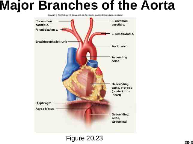
Major Branches of the Aorta Copyright The McGraw-Hill Companies, Inc. Permission required for reproduction or display. L. common carotid a. R. common carotid a. R. subclavian a. L. subclavian a. Brachiocephalic trunk Aortic arch Ascending aorta Descending aorta, thoracic (posterior to heart) Diaphragm Aortic hiatus Descending aorta, abdominal Figure 20.23 20-3
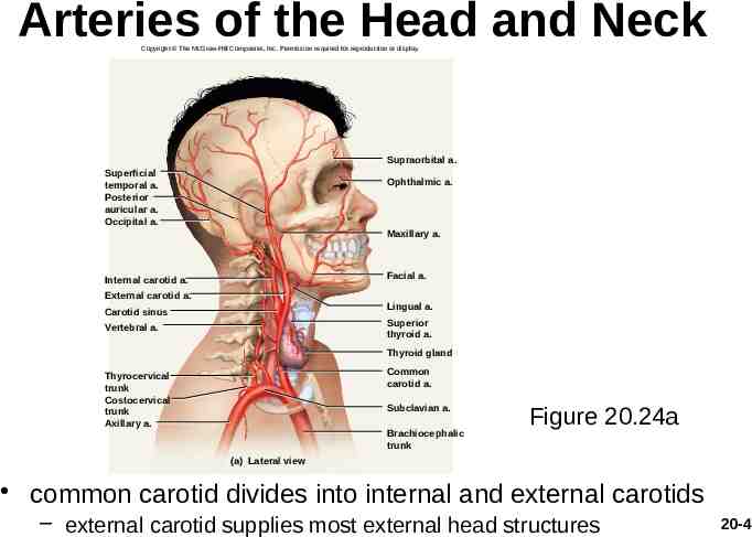
Arteries of the Head and Neck Copyright The McGraw-Hill Companies, Inc. Permission required for reproduction or display. Supraorbital a. Superficial temporal a. Posterior auricular a. Occipital a. Ophthalmic a. Maxillary a. Facial a. Internal carotid a. External carotid a. Lingual a. Carotid sinus Superior thyroid a. Vertebral a. Thyroid gland Common carotid a. Thyrocervical trunk Costocervical trunk Axillary a. Subclavian a. Brachiocephalic trunk Figure 20.24a (a) Lateral view common carotid divides into internal and external carotids – external carotid supplies most external head structures 20-4
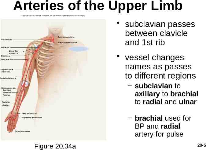
Arteries of the Upper Limb Copyright The McGraw-Hill Companies, Inc. Permission required for reproduction or display. Common carotid a. Subclavian a. Brachiocephalic trunk Axillary a. Circumflex humeral aa. Brachial a. subclavian passes between clavicle and 1st rib vessel changes names as passes to different regions Deep brachial a. Superior ulnar collateral a. Radial collateral a. – subclavian to axillary to brachial to radial and ulnar Interosseous aa.: Common Posterior Anterior Radial a. Ulnar a. Deep palmar arch Superficial palmar arch (a) Major arteries Figure 20.34a – brachial used for BP and radial artery for pulse 20-5
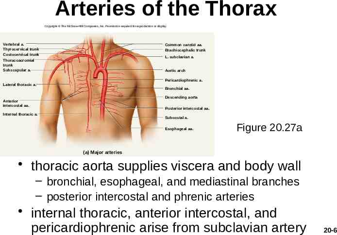
Arteries of the Thorax Copyright The McGraw-Hill Companies, Inc. Permission required for reproduction or display. Vertebral a. Thyrocervical trunk Costocervical trunk Common carotid aa. Brachiocephalic trunk L. subclavian a. Thoracoacromial trunk Subscapular a. Aortic arch Pericardiophrenic a. Lateral thoracic a. Bronchial aa. Descending aorta Anterior intercostal aa. Posterior intercostal aa. Internal thoracic a. Subcostal a. Esophageal aa. Figure 20.27a (a) Major arteries thoracic aorta supplies viscera and body wall – bronchial, esophageal, and mediastinal branches – posterior intercostal and phrenic arteries internal thoracic, anterior intercostal, and pericardiophrenic arise from subclavian artery 20-6
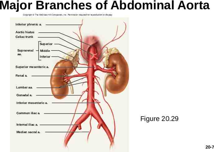
Major Branches of Abdominal Aorta Copyright The McGraw-Hill Companies, Inc. Permission required for reproduction or display. Inferior phrenic a. Aortic hiatus Celiac trunk Superior Suprarenal aa. Middle Inferior Superior mesenteric a. Renal a. Lumbar aa. Gonadal a. Inferior mesenteric a. Common iliac a. Figure 20.29 Internal iliac a. Median sacral a. 20-7
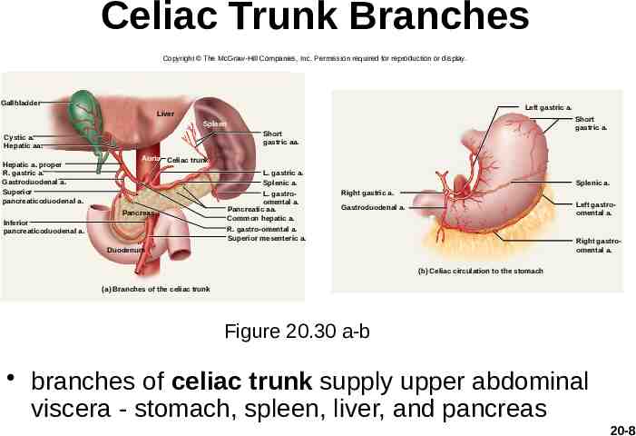
Celiac Trunk Branches Copyright The McGraw-Hill Companies, Inc. Permission required for reproduction or display. Gallbladder Left gastric a. Liver Short gastric a. Spleen Short gastric aa. Cystic a. Hepatic aa. Hepatic a. proper R. gastric a. Gastroduodenal a. Superior pancreaticoduodenal a. Aorta Celiac trunk Pancreas Inferior pancreaticoduodenal a. L. gastric a. Splenic a. L. gastroomental a. Pancreatic aa. Common hepatic a. R. gastro-omental a. Superior mesenteric a. Splenic a. Right gastric a. Left gastroomental a. Gastroduodenal a. Right gastroomental a. Duodenum (b) Celiac circulation to the stomach (a) Branches of the celiac trunk Figure 20.30 a-b branches of celiac trunk supply upper abdominal viscera - stomach, spleen, liver, and pancreas 20-8
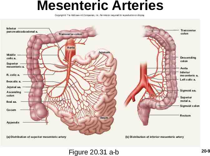
Mesenteric Arteries Copyright The McGraw-Hill Companies, Inc. Permission required for reproduction or display. Inferior pancreaticoduodenal a. Transverse colon Transverse colon Aorta Middle colic a. Jejunum Descending colon Superior mesenteric a. Aorta Inferior mesenteric a. Left colic a. R. colic a. Ileocolic a. Jejunal aa. Sigmoid aa. Ascending colon Superior rectal a. Ileal aa. Sigmoid colon Cecum Ileum Rectum Appendix (a) Distribution of superior mesenteric artery (b) Distribution of inferior mesenteric artery Figure 20.31 a-b 20-9
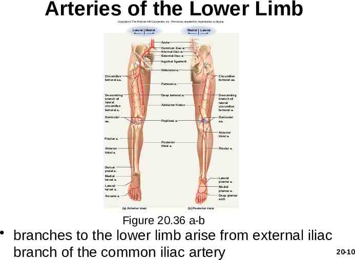
Arteries of the Lower Limb Copyright The McGraw-Hill Companies, Inc. Permission required for reproduction or display. Lateral Medial Medial Lateral Aorta Common iliac a. Internal iliac a. External iliac a. Inguinal ligament Obturator a. Circumflex femoral aa. Circumflex femoral aa. Femoral a. Descending branch of lateral circumflex femoral a. Genicular aa. Deep femoral a. Descending branch of lateral circumflex femoral a. Adductor hiatus Genicular aa. Popliteal a. Anterior tibial a. Fibular a. Posterior tibial a. Anterior tibial a. Fibular a. Dorsal pedal a. Medial tarsal a. Lateral plantar a. Lateral tarsal a. Medial plantar a. Arcuate a. Deep plantar arch (a) Anterior view (b) Posterior view Figure 20.36 a-b branches to the lower limb arise from external iliac 20-10 branch of the common iliac artery
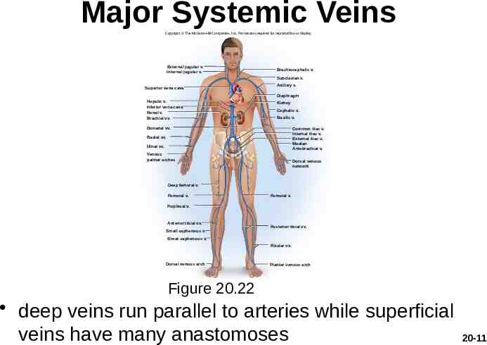
Major Systemic Veins Copyright The McGraw-Hill Companies, Inc. Permission required for reproduction or display. External jugular v. Internal jugular v. Brachiocephalic v. Subclavian v. Superior vena cava Axillary v. Diaphragm Hepatic v. Inferior vena cava Renal v. Brachial vv. Kidney Cephalic v. Basilic v. Gonadal vv. Common iliac v. Internal iliac v. External iliac v. Median Antebrachial v. Radial vv. Ulnar vv. Venous palmar arches Dorsal venous network Deep femoral v. Femoral v. Femoral v. Popliteal v. Anterior tibial vv. Small saphenous v. Posterior tibial vv. Great saphenous v. Fibular vv. Dorsal venous arch Plantar venous arch Figure 20.22 deep veins run parallel to arteries while superficial veins have many anastomoses 20-11
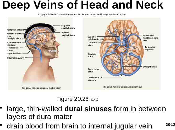
Deep Veins of Head and Neck Copyright The McGraw-Hill Companies, Inc. Permission required for reproduction or display. Superior sagittal sinus Corpus callosum Inferior sagittal sinus Great cerebral vein Straight sinus Superior ophthalmic vein Confluence of sinuses Transverse sinus Cavernous sinus Sigmoid sinus Sigmoid sinus Internal jugularv. Superficial middle cerebral vein To internal jugular v. Straight sinus Transverse sinus Confluence of sinuses (a) Dural venous sinuses, medial view (b) Dural venous sinuses, inferior view Figure 20.26 a-b large, thin-walled dural sinuses form in between layers of dura mater 20-12 drain blood from brain to internal jugular vein
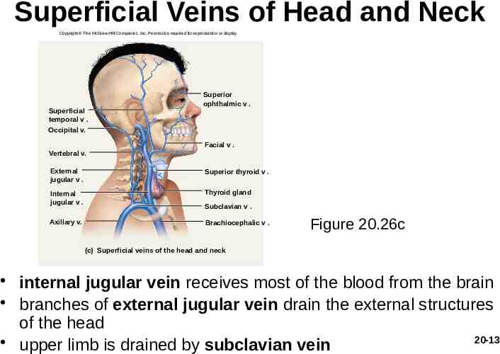
Superficial Veins of Head and Neck Copyright The McGraw-Hill Companies, Inc. Permission required for reproduction or display. Superficial temporal v . Occipital v. Superior ophthalmic v . Facial v . Vertebral v. External jugular v . Superior thyroid v . Internal jugular v . Thyroid gland Axillary v. Brachiocephalic v . Subclavian v . Figure 20.26c (c) Superficial veins of the head and neck internal jugular vein receives most of the blood from the brain branches of external jugular vein drain the external structures of the head 20-13 upper limb is drained by subclavian vein
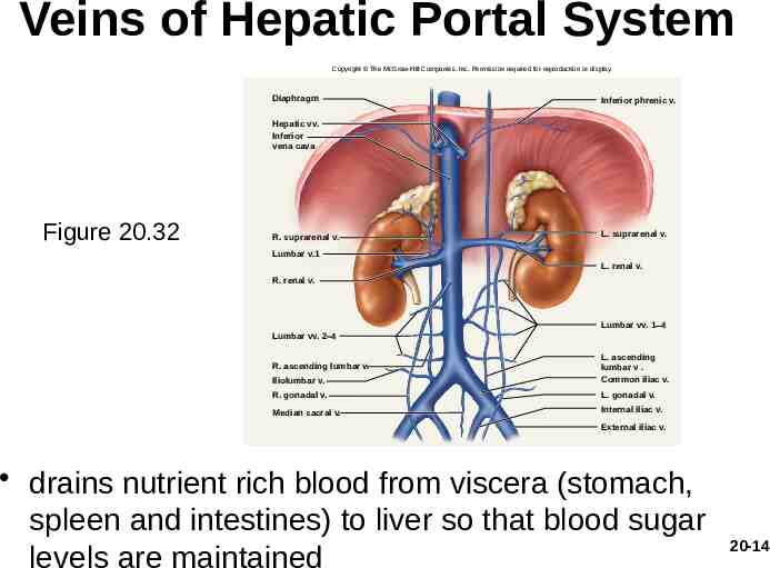
Veins of Hepatic Portal System Copyright The McGraw-Hill Companies, Inc. Permission required for reproduction or display. Diaphragm Inferior phrenic v. Hepatic vv. Inferior vena cava Figure 20.32 R. suprarenal v. L. suprarenal v. Lumbar v.1 L. renal v. R. renal v. Lumbar vv. 1-4 Lumbar vv. 2–4 R. ascending lumbar v. Iliolumbar v. L. ascending lumbar v . Common iliac v. R. gonadal v. L. gonadal v. Median sacral v. Internal iliac v. External iliac v. drains nutrient rich blood from viscera (stomach, spleen and intestines) to liver so that blood sugar levels are maintained 20-14
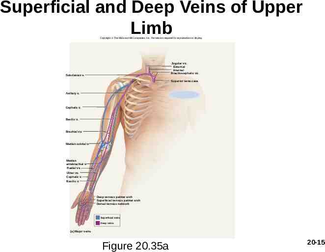
Superficial and Deep Veins of Upper Limb Copyright The McGraw-Hill Companies, Inc. Permission required for reproduction or display. Jugular vv. External Internal Brachiocephalic vv. Subclavian v. Superior vena cava Axillary v. Cephalic v. Basilic v. Brachial vv. Median cubital v. Median antebrachial v. Radial vv. Ulnar vv. Cephalic v. Basilic v. Deep venous palmar arch Superficial venous palmar arch Dorsal venous network Superficial veins Deep veins (a) Major veins Figure 20.35a 20-15
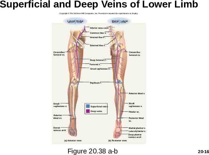
Superficial and Deep Veins of Lower Limb Copyright The McGraw-Hill Companies, Inc. Permission required for reproduction or display. Lateral Medial Medial Lateral Inferior vena cava Common iliac v. Internal iliac v . External iliac v . Circumflex femoral vv. Circumflex femoral vv. Deep femoral v . Femoral v . Great saphenous v . Popliteal v . Anterior tibial v. Small saphenous v. Superficial veins Small saphenous v. Deep veins Fibular vv. Anterior tibial vv. Posterior tibial vv. Dorsal venous arch Medial plantar v. Lateral plantar v. Deep plantar venous arch (a) Anterior view (b) Posterior view Figure 20.38 a-b 20-16
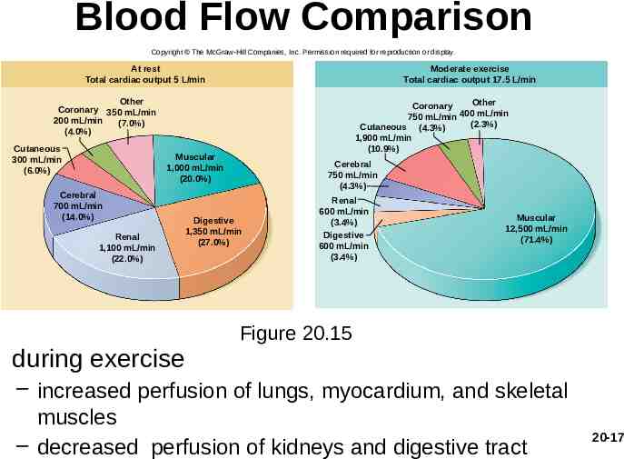
Blood Flow Comparison Copyright The McGraw-Hill Companies, Inc. Permission required for reproduction or display. At rest Total cardiac output 5 L/min Moderate exercise Total cardiac output 17.5 L/min Other Coronary 350 mL/min 200 mL/min (7.0%) (4.0%) Cutaneous 300 mL/min (6.0%) Other Coronary 400 mL/min 750 mL/min (2.3%) Cutaneous (4.3%) 1,900 mL/min (10.9%) Muscular 1,000 mL/min (20.0%) Cerebral 700 mL/min (14.0%) Renal 1,100 mL/min (22.0%) Cerebral 750 mL/min (4.3%) Digestive 1,350 mL/min (27.0%) Renal 600 mL/min (3.4%) Digestive 600 mL/min (3.4%) Muscular 12,500 mL/min (71.4%) Figure 20.15 during exercise – increased perfusion of lungs, myocardium, and skeletal muscles – decreased perfusion of kidneys and digestive tract 20-17
What is blood pressure? Measures force of blood in the arteries High blood pressure (HBP) hypertension (HTN)
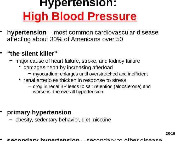
Hypertension: High Blood Pressure hypertension – most common cardiovascular disease affecting about 30% of Americans over 50 “the silent killer” – major cause of heart failure, stroke, and kidney failure damages heart by increasing afterload – myocardium enlarges until overstretched and inefficient renal arterioles thicken in response to stress – drop in renal BP leads to salt retention (aldosterone) and worsens the overall hypertension primary hypertension – obesity, sedentary behavior, diet, nicotine 20-19
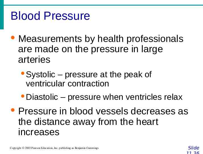
Blood Pressure · Measurements by health professionals are made on the pressure in large arteries · Systolic – pressure at the peak of ventricular contraction · Diastolic – pressure when ventricles relax · Pressure in blood vessels decreases as the distance away from the heart increases Copyright 2003 Pearson Education, Inc. publishing as Benjamin Cummings Slide
Blood Pressure: Effects of Factors · Neural factors · Autonomic nervous system adjustments (sympathetic division) and parasympathetic · Renal factors · Regulation by altering blood volume · Renin – hormonal control Copyright 2003 Pearson Education, Inc. publishing as Benjamin Cummings Slide
Blood Pressure: Effects of Factors · Temperature · Heat has a vasodilation effect · Cold has a vasoconstricting effect · Chemicals · Various substances can cause increases or decreases · Endocrine/hormone functions as stimulators or relaxors. · Diet Copyright 2003 Pearson Education, Inc. publishing as Benjamin Cummings Slide
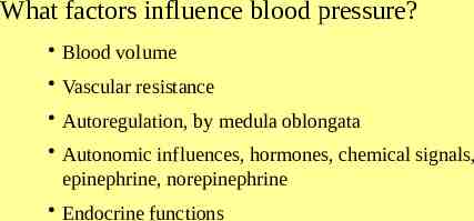
What factors influence blood pressure? Blood volume Vascular resistance Autoregulation, by medula oblongata Autonomic influences, hormones, chemical signals, epinephrine, norepinephrine Endocrine functions
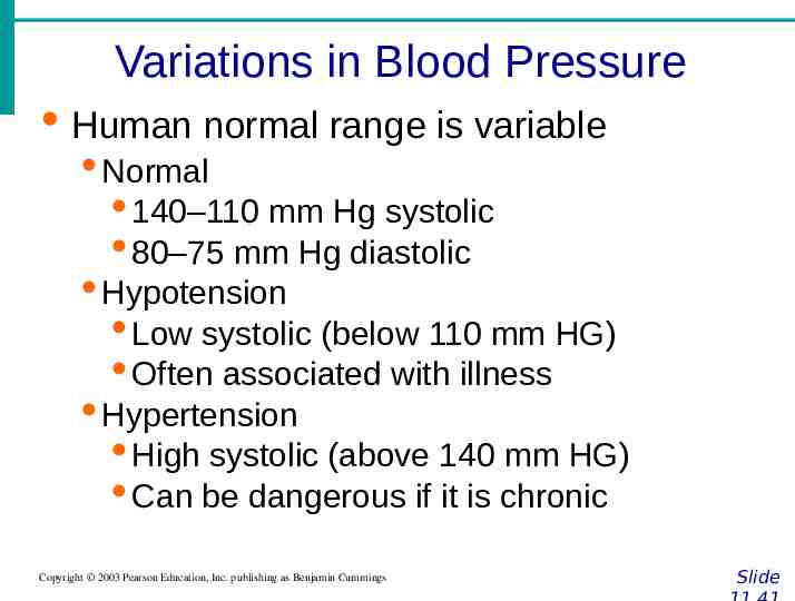
Variations in Blood Pressure · Human normal range is variable · Normal · 140–110 mm Hg systolic · 80–75 mm Hg diastolic · Hypotension · Low systolic (below 110 mm HG) · Often associated with illness · Hypertension · High systolic (above 140 mm HG) · Can be dangerous if it is chronic Copyright 2003 Pearson Education, Inc. publishing as Benjamin Cummings Slide
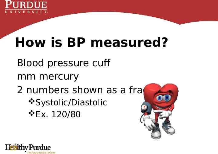
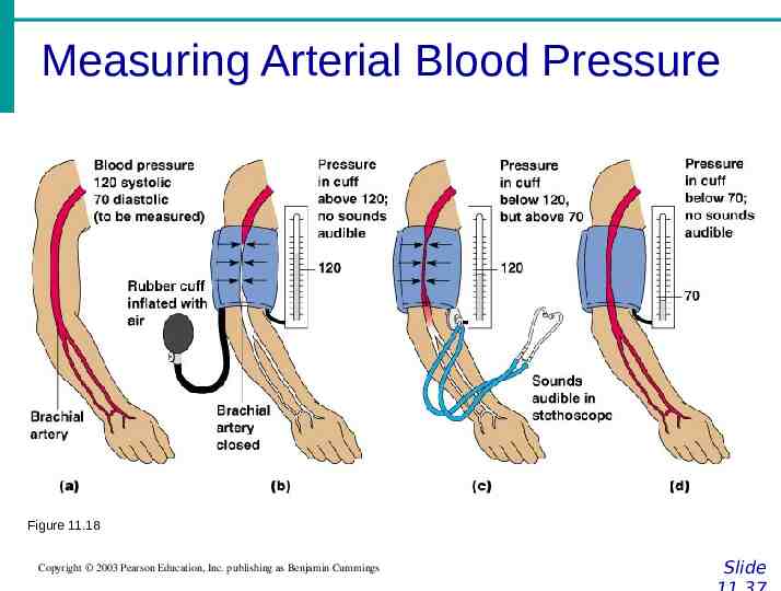
How is BP measured? Blood pressure cuff mm mercury 2 numbers shown as a fraction Systolic/Diastolic Ex. 120/80
Systolic The top number in blood pressure readings Measures the pressure in arteries when heart is beating
Diastolic The bottom number in blood pressure readings Measures pressure when heart is at rest
Measuring Arterial Blood Pressure Figure 11.18 Copyright 2003 Pearson Education, Inc. publishing as Benjamin Cummings Slide
How to take your blood pressure https://www.youtube.com/watch?v Gmic13m vsgo