Development and Growth of Teeth:
40 Slides3.61 MB
Development and Growth of Teeth:
The primitive oral cavity, or stomodeum, is lined by stratified squamous epithelium called the oral ectoderm or primitive oral epithelium. The oral ectoderm contacts the endoderm of the foregut to form the buccopharyngeal membrane. At about the twenty-seventh day of gestation this membrane ruptures and the primitive oral cavity establishes a connection with the foregut.
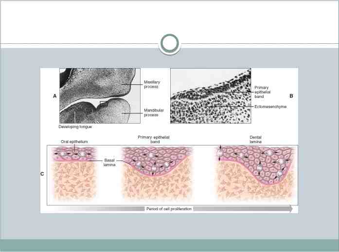
Most of the connective tissue cells underlying the oral ectoderm are of neural crest or ectomesenchyme in origin. These cells are thought to instruct or induce the overlying ectoderm to start tooth development, which begins in the anterior portion of what will be the future maxilla and mandible and proceeds posteriorly
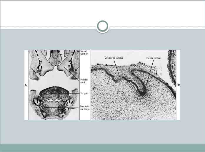
DENTAL LAMINA: Two or 3 weeks after the rupture of the buccopharyngeal membrane, when the embryo is about 6 weeks old, certain areas of basal cells of the oral ectoderm proliferate more rapidly than do the cells of the adjacent areas. This leads to the formation of the Primary epithelial band which is a band of epithelium that has invaded the underlying ectomesenchyme along each of the horseshoe-shaped future dental arches .
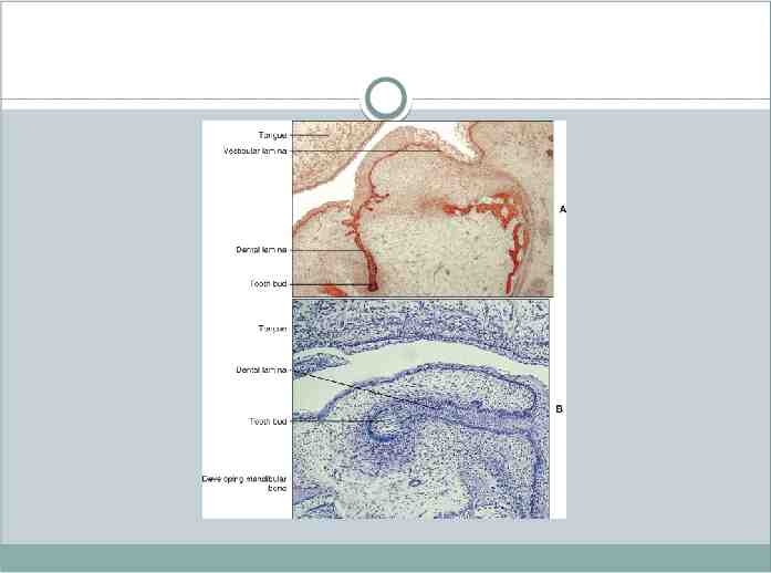
At about 7th week the primary epithelial band divides into an inner (lingual) process called Dental lamina and an outer (buccal) process called Vestibular lamina. The dental laminae serve as the primordium for the ectodermal portion of the deciduous teeth. Later, during the development of the jaws, the permanent molars arise directly from a distal extension of the dental lamina.
The development of the first permanent molar is initiated at the fourth month in utero. The second molar is initiated at about the first year after birth, the third molar at the fourth or fifth years. The distal proliferation of the dental lamina is responsible for the location of the germs of the permanent molars in the ramus of the mandible and the tuberosity of the maxilla.
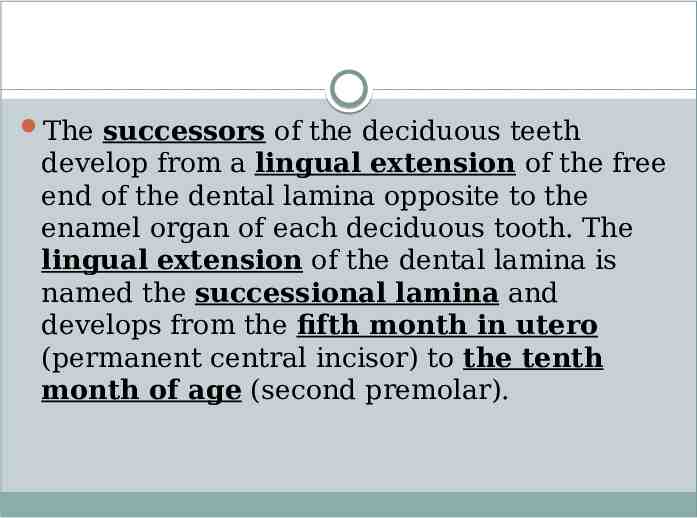
The successors of the deciduous teeth develop from a lingual extension of the free end of the dental lamina opposite to the enamel organ of each deciduous tooth. The lingual extension of the dental lamina is named the successional lamina and develops from the fifth month in utero (permanent central incisor) to the tenth month of age (second premolar).
Fate of dental lamina: It is evident that the total activity of the dental lamina extends over a period of at least 5 years. Any particular portion of the dental lamina functions for a much briefer period since only a relatively short time elapses after initiation of tooth development before the dental lamina begins to degenerate at that particular location. However, the dental lamina may still be active in the third molar region after it has disappeared elsewhere, except for occasional epithelial remnants.
As the teeth continue to develop, they lose their connection with the dental lamina. They later break up by mesenchymal invasion, which is at first incomplete and does not perforate the total thickness of the lamina. Remnants of the dental lamina persist as epithelial pearls or islands within the jaw as well as in the gingiva. These are referred to as cell rest of Serres.
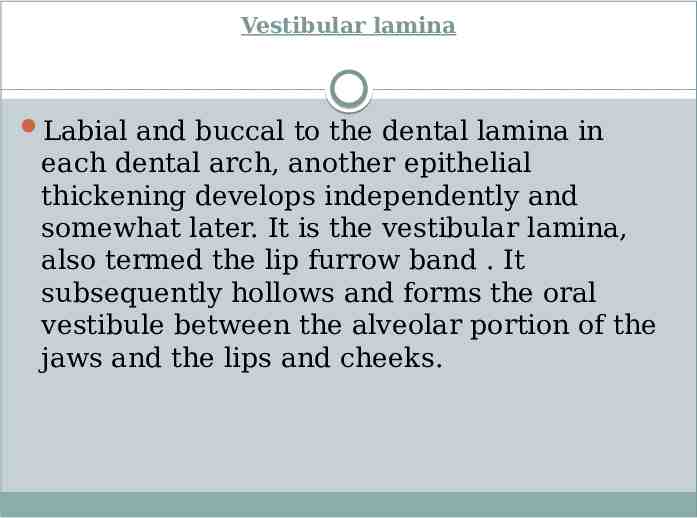
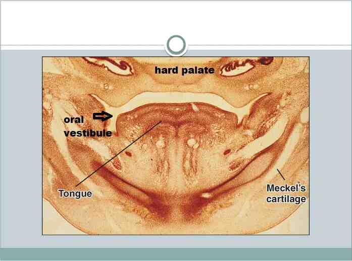
Vestibular lamina Labial and buccal to the dental lamina in each dental arch, another epithelial thickening develops independently and somewhat later. It is the vestibular lamina, also termed the lip furrow band . It subsequently hollows and forms the oral vestibule between the alveolar portion of the jaws and the lips and cheeks.
TOOTH DEVELOPMENT: At certain points along the dental lamina, each representing the location of one of the 10 mandibular and 10 maxillary deciduous teeth, the ectodermal cells multiply still more rapidly and form little knobs that grow into the underlying mes- enchyme . Each of these little downgrowths from the dental lamina represents the beginning of the enamel organ of the tooth bud of a deciduous tooth. Not all of these enamel organs start to develop at the same time, and the first to appear are those of the anterior mandibular region.
As cell proliferation continues, each enamel organ increases in size, sinks deeper into the ectomesenchyme and due to differential growth changes its shape. As it develops, it takes on a shape that resembles a cap, with an outer convex surface facing the oral cavity and an inner concavity. On the inside of the cap (i.e., inside the depression of the enamel organ), the ectomesenchymal cells increase in number. The tissue appears more dense than the surrounding mesenchyme and represents the beginning of the dental papilla.
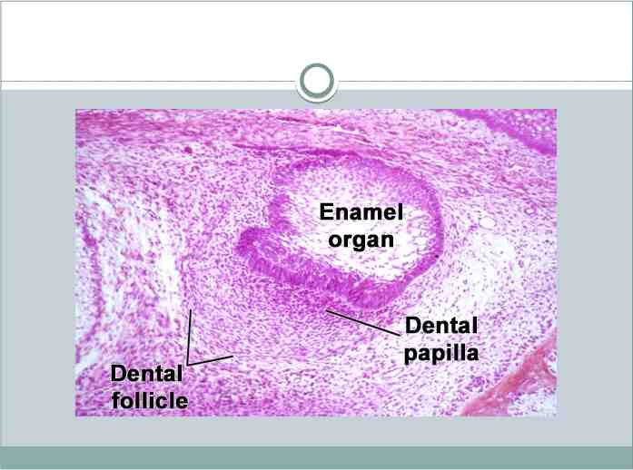
Surrounding the combined enamel organ and dental papilla, the third part of the tooth bud forms. It is the dental sac or dental follicle, and it consists of ectomesenchymal cells and fibers that surround the dental papilla and the enamel organ. Thus the tooth germ consists of the ectodermal
component—the enamel organ and the ectomesenchymal components—the dental papilla and the dental follicle. The tooth and its supporting structures are formed from the tooth germ. The enamel is formed from the enamel organ, the dentin and pulp from the dental papilla and the supporting tissues namely the cementum, periodontal ligament and the alveolar bone from the dental follicle.
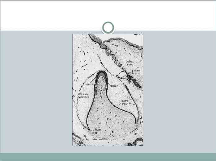
During and after these developments the shape of the enamel organ continues to change. The depression occupied by the dental papilla deepens until the enamel organ assumes a shape resembling a bell. As this development takes place, the dental lamina, which had thus far connected the enamel organ to the oral epithelium, becomes longer and thinner and finally breaks up and the tooth bud loses its connection with the epithelium of the primitive oral cavity. Development of tooth results from interaction of the epithelium derived from the first arch and ectomesenchymal cells derived from the neural crest cells. Up to 12 days the first arch.
Like any other organ development in our body numerous and complex gene expression occurs to control the development process through molecular signals. In odontogenesis, many of the genes involved or the molecular signals directed by them are common to other developing organs like kidney and lung or structures like the limb.
DEVELOPMENTAL STAGES Although tooth development is a continuous process, the developmental history of a tooth is divided into several morphologic “stages” for descriptive purposes. While the size and shape of individual teeth are different, they pass through similar stages of development. They are named after the shape of the enamel organ (epithelial part of the tooth germ), and are called the bud, cap, and bell stages.
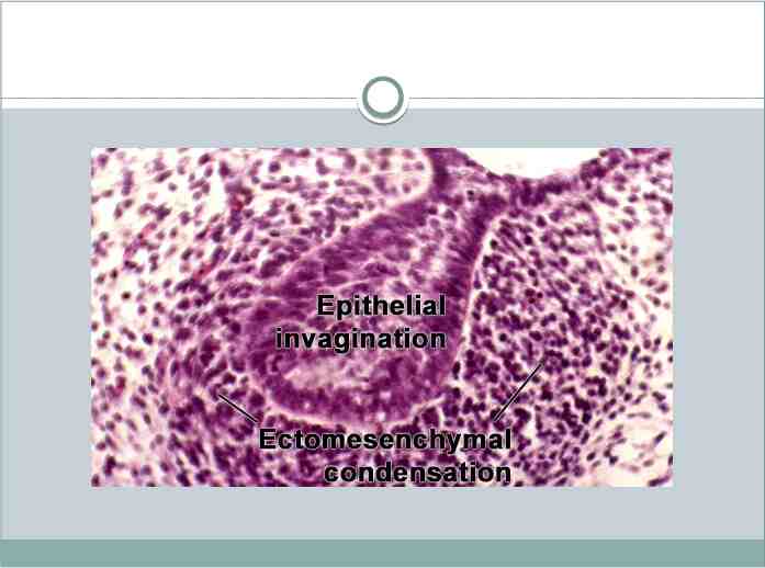
1-Bud stage: The epithelium of the dental laminae is separated from the underlying ectomesenchyme by a basement membrane. Simultaneous with the differentiation of each dental lamina, round or ovoid swellings arise from the basement membrane at 10 different points, corresponding to the future positions of the deciduous teeth. These are the primordia of the enamel organs, the tooth buds. Thus the development of tooth germs is initiated, and the cells continue to proliferate faster than adjacent cells.
The dental lamina is shallow, and microscopic sections often show tooth buds close to the oral epithelium. Since the main function of certain epithelial cells of the tooth bud is to form the tooth enamel, these cells constitute the enamel organ, which is critical to normal tooth development. In the bud stage, the enamel organ consists of peripherally located low columnar cells and centrally located polygonal cells. Many cells of the tooth bud and the surrounding mesenchyme undergo mitosis.
As a result of the increased mitotic activity and the migration of neural crest cells into the area the ectomesenchymal cells surrounding the tooth bud condense. The area of ectomesenchymal condensation immediately subjacent to the enamel organ is the dental papilla. The condensed ectomesenchyme that surrounds the tooth bud and the dental papilla is the dental sac . Both the dental papilla and the dental sac become more well defined as the enamel organ grows into the cap and bell shapes .
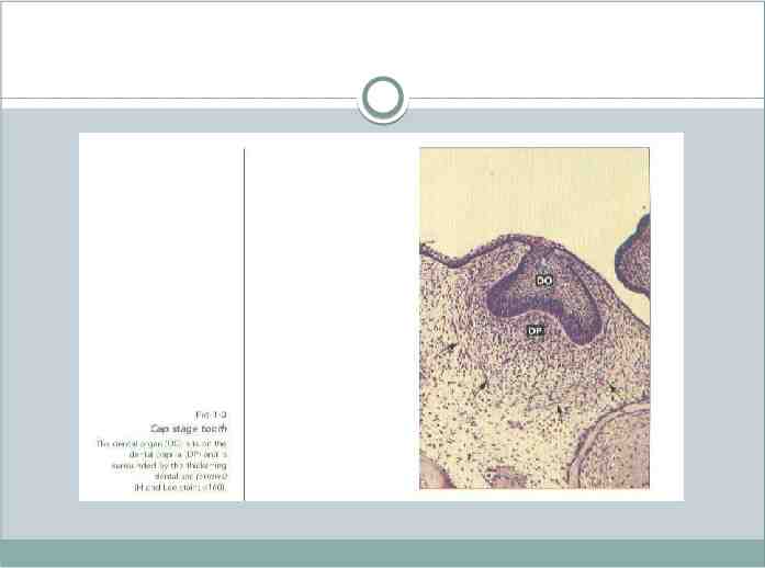
Cap stage: As the tooth bud continues to proliferate, it does not expand uniformly into a larger sphere. Instead, unequal growth in different parts of the tooth bud leads to the cap stage, which is characterized by a shallow invagination on the deep surface of the bud
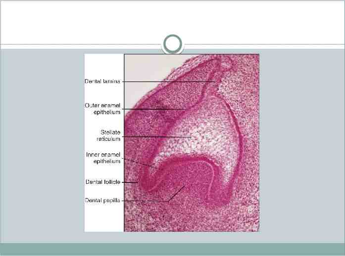
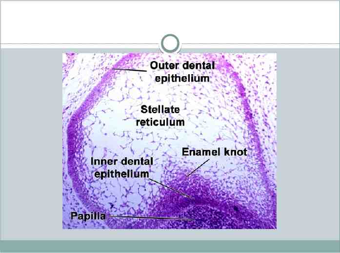
Outer and inner enamel epithelium The peripheral cells of the cap stage are cuboidal, cover the convexity of the “cap,” and are called the outer enamel (dental) epithelium. The cells in the concavity of the “cap” become tall, columnar cells and represent the inner enamel (dental) epithelium . The outer enamel epithelium is separated from the dental sac, and the inner enamel epithelium from the dental papilla, by a delicate basement membrane.
Hemidesmosomes anchor the cells to the basal lamina. The enamel organ may be seen to have a double attachment of dental lamina to the overlying oral epithelium enclosing ectomesenchyme called enamel niche between them. This appearance is due to a funnel-shaped depression of the dental lamina.
Stellate reticulum: Polygonal cells located in the center of the epithelial enamel organ, between the outer and inner enamel epithelia, begin to separate due to water being drawn into the enamel organ from the surrounding dental papilla as a result of osmotic force exerted by glycosaminoglycans contained in the ground substance. As a result the polygonal cells become star shaped but maintain contact with each other by their cytoplasmic process. As these star- shaped cells form a cellular network, they are called the stellate reticulum .
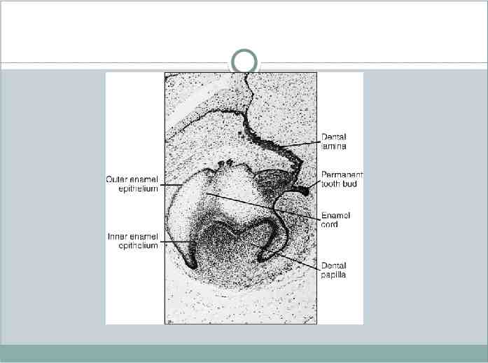
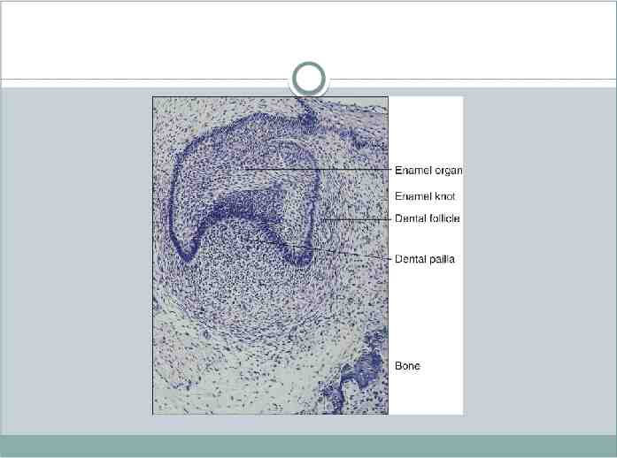
This gives the stellate reticulum a cushion like consistency and acts as a shock absorber that may support and protect the delicate enamel-forming cells. The cells in the center of the enamel organ are densely packed and form the enamel knot. This knot projects in part toward the underlying dental papilla, so that the center of the epithelial invagination shows a slightly knob like enlargement that is bordered by the labial and lingual enamel grooves.At the same time a vertical extension of the enamel knot, called the enamel cord occurs .
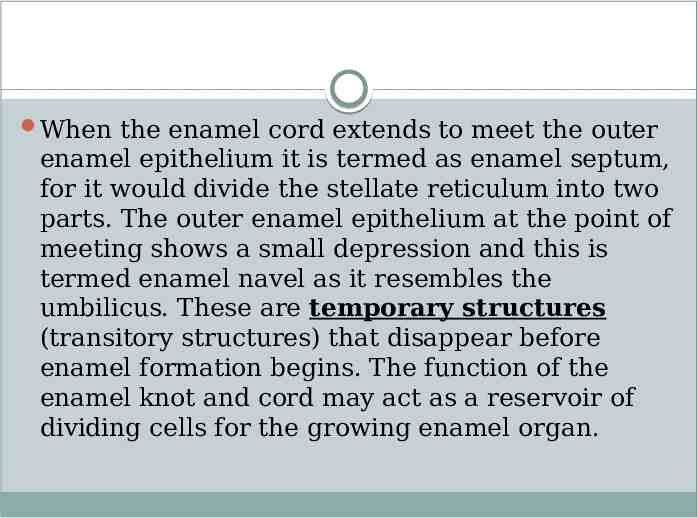
When the enamel cord extends to meet the outer enamel epithelium it is termed as enamel septum, for it would divide the stellate reticulum into two parts. The outer enamel epithelium at the point of meeting shows a small depression and this is termed enamel navel as it resembles the umbilicus. These are temporary structures (transitory structures) that disappear before enamel formation begins. The function of the enamel knot and cord may act as a reservoir of dividing cells for the growing enamel organ.
Recent studies have shown that enamel knot acts as a signaling center as many important growth factors are expressed by the cells of the enamel knot and thus they play an important part in determining the shape of the tooth. These are discussed in detail in the section on molecular insights in tooth morphogenesis.