PREVENTION AND TREATMENT OF PRESSURE ULCERS By Holly Ferguson, PT, WCC


66 Slides1.46 MB
PREVENTION AND TREATMENT OF PRESSURE ULCERS By Holly Ferguson, PT, WCC
PURPOSE The purpose of this self-learning module is to help guide nurses performing skin assessment and care according to best practice. At the end of the module, the nurse will be able to: Identify skin assessment protocol Identify risk factors for developing pressure ulcers Identify at least 3 things to help prevent pressure ulcers Identify the SJH nursing and interdisciplinary team resources Identify ways to manage and treat pressure ulcers
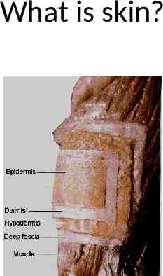
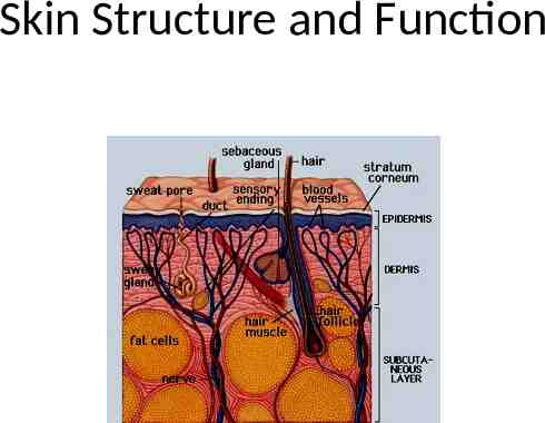
What is skin?
Skin Structure and Function
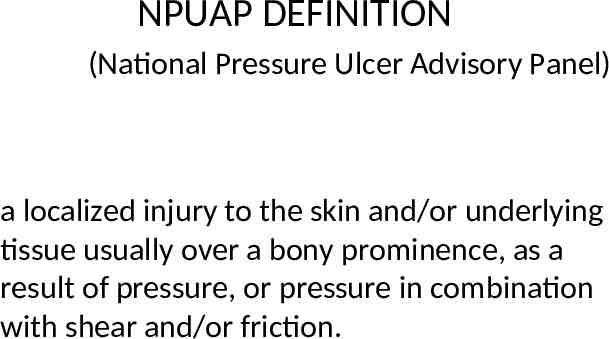
NPUAP DEFINITION (National Pressure Ulcer Advisory Panel) a localized injury to the skin and/or underlying tissue usually over a bony prominence, as a result of pressure, or pressure in combination with shear and/or friction.
PRESSURE ON VESSELS Unrelieved pressure on the skin squeezes tiny blood vessels, which supply the skin with nutrients and oxygen. When the skin is starved for too long, the tissue dies, and a pressure ulcer develops
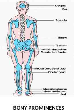
BONY PROMINENCES
COMMON SITES
CLINICAL PRESENTATION 1. Rounded, crater like shapes with regular edges 2. Over bony prominences, but can take on the shape of the bone (malleoli vs sacrum) 3. Usually dark regular base that do not bleed easily 4. 95% over sacrum/coccyx, trochanter, IT, heel and lateral malleoli
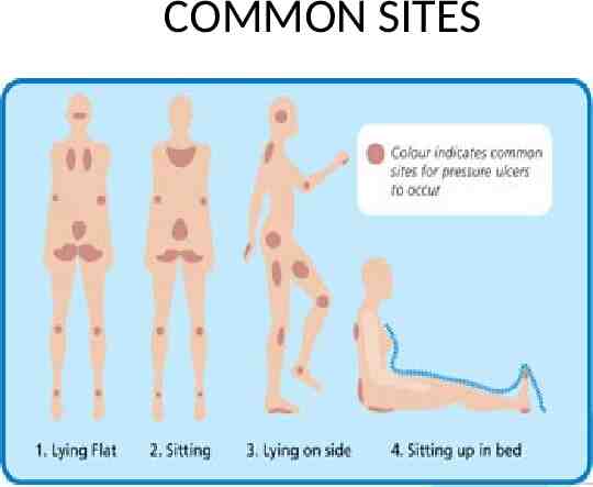
STATISTICS 70% occur in people over 65 Most common sites SACRUM AND HEELS Shoulder, heel, and ear were the favorite sites of newly developed PrUs
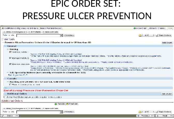
EPIC ORDER SET: PRESSURE ULCER PREVENTION
STAGES/PROGRESSION 4 stages depending on amount and time of ischemia 1.Hyperemia (redness) will occur within 30 minutes of sustained pressure. If the Pressure is removed the hyperemia will disappear in approximately 24-36 hours 2.Depending on a number factors (including general health) ischemia begins in 2-6 hours of sustained pressure 3.Texture usually feels hard 4.Necrosis begins 6 hours after sustained pressure. 5.At this stage skin appears blue or greyish and may be indurated. Recovery at this stage is variable. 6.Finally, if pressure continues, ulceration occurs.
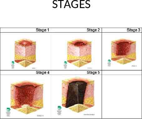
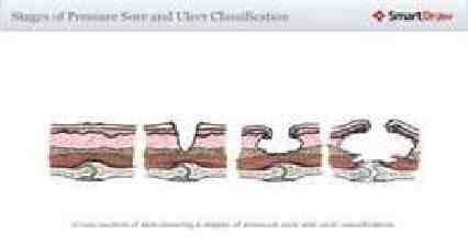
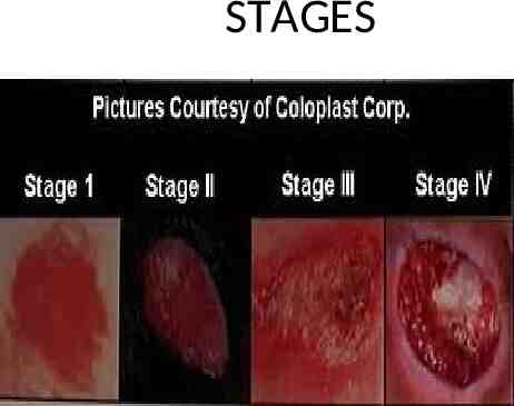
STAGES
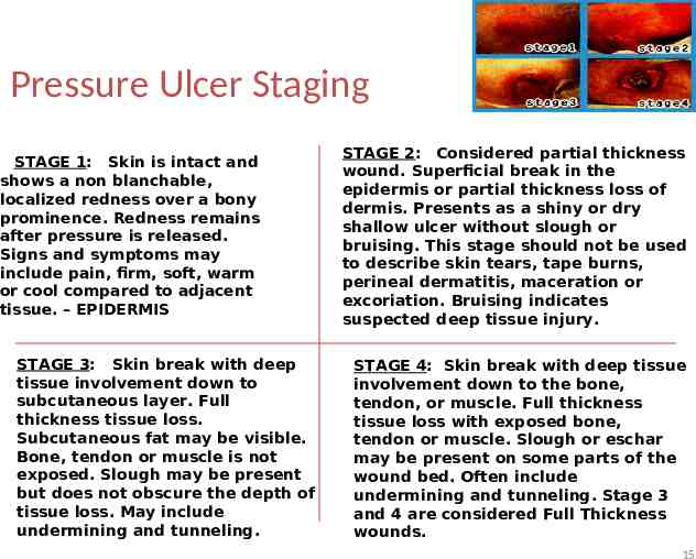
Pressure Ulcer Staging STAGE 1: Skin is intact and shows a non blanchable, localized redness over a bony prominence. Redness remains after pressure is released. Signs and symptoms may include pain, firm, soft, warm or cool compared to adjacent tissue. – EPIDERMIS STAGE 3: Skin break with deep tissue involvement down to subcutaneous layer. Full thickness tissue loss. Subcutaneous fat may be visible. Bone, tendon or muscle is not exposed. Slough may be present but does not obscure the depth of tissue loss. May include undermining and tunneling. STAGE 2: Considered partial thickness wound. Superficial break in the epidermis or partial thickness loss of dermis. Presents as a shiny or dry shallow ulcer without slough or bruising. This stage should not be used to describe skin tears, tape burns, perineal dermatitis, maceration or excoriation. Bruising indicates suspected deep tissue injury. STAGE 4: Skin break with deep tissue involvement down to the bone, tendon, or muscle. Full thickness tissue loss with exposed bone, tendon or muscle. Slough or eschar may be present on some parts of the wound bed. Often include undermining and tunneling. Stage 3 and 4 are considered Full Thickness wounds. 15
STAGES
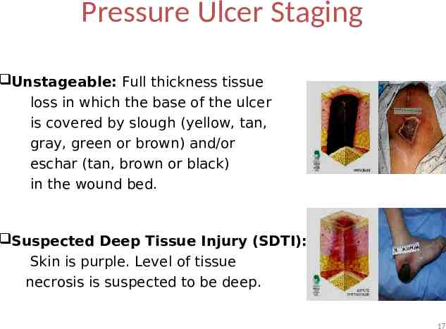
Pressure Ulcer Staging Unstageable: Full thickness tissue loss in which the base of the ulcer is covered by slough (yellow, tan, gray, green or brown) and/or eschar (tan, brown or black) in the wound bed. Suspected Deep Tissue Injury (SDTI): Skin is purple. Level of tissue necrosis is suspected to be deep. 17
*difference between stage II and stage III – stage II will never have slough *Never back stage a PrU; document a “healing stage III” not a “stage II”
HEELS Second most common site for PrU’s, and most common site of DTI Color reflects the degree of DTI: – Red - hyperemia and ischemia – Purple – infarction – Black - necrosis
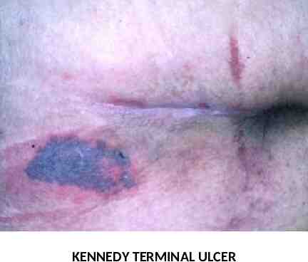
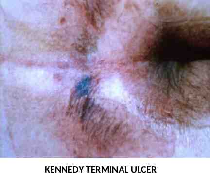
UNAVOIDABLE PRESSURE ULCERS According to NPUAP: An individual develops a PrU even thought the provider had evaluated the individual’s clinical condition and pressure ulcer risk factors’ defined and implemented interventions that are consistent with individual needs, goals and recognized standards practice; monitored and evaluated the impact of the interventions; and revised the approaches as appropriate “Kennedy Terminal Ulcers”
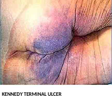
KENNEDY TERMINAL ULCERS Ulcers that some people get as they are dying Usually on the sacrum Usually pear shaped Colors vary Irregular borders Come on suddenly Geriatric phenomenon
KENNEDY TERMINAL ULCERS CONTINUED Often confused for dirt or dried stool in the beginning, and providers try to wash it off only to find it is under the skin In the beginning it can look like the skin got scraped off in a bad abrasion Progresses rapidly (over a matter of hours)
KENNEDY TERMINAL ULCER
KENNEDY TERMINAL ULCER
KENNEDY TERMINAL ULCER
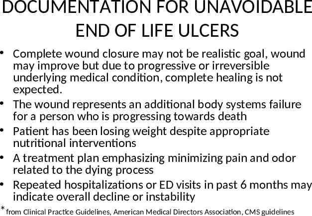
DOCUMENTATION FOR UNAVOIDABLE END OF LIFE ULCERS Complete wound closure may not be realistic goal, wound may improve but due to progressive or irreversible underlying medical condition, complete healing is not expected. The wound represents an additional body systems failure for a person who is progressing towards death Patient has been losing weight despite appropriate nutritional interventions A treatment plan emphasizing minimizing pain and odor related to the dying process Repeated hospitalizations or ED visits in past 6 months may indicate overall decline or instability *from Clinical Practice Guidelines, American Medical Directors Association, CMS guidelines
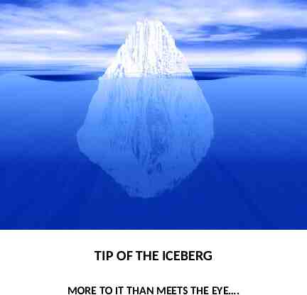
TIP OF THE ICEBERG MORE TO IT THAN MEETS THE EYE .
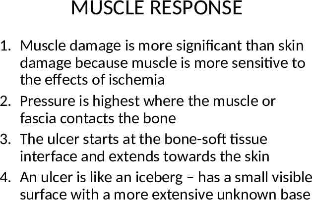
MUSCLE RESPONSE 1. Muscle damage is more significant than skin damage because muscle is more sensitive to the effects of ischemia 2. Pressure is highest where the muscle or fascia contacts the bone 3. The ulcer starts at the bone-soft tissue interface and extends towards the skin 4. An ulcer is like an iceberg – has a small visible surface with a more extensive unknown base
“Assess the whole person; not just the hole in the person”. Gary Sibbold 29
EXTRINSIC FACTORS
FRICTION 1. Rubbing 2 surfaces against another 2. Friction without pressure causes damage to only the epidermis and upper dermal layer (think sheet burn)
SHEAR 1. Friction Gravity Shear 2. Think elevation of head of bed and person sliding down in bed or sliding down in reclining chair 3. Shear stretches and tears vessels, which reduces the amount of pressure necessary to cause ischemia and deep tissue injury 4. Shearing can cause undermining and tunneling *blisters
DRYNESS Stratum corneum normally has 10-15% moisture When the moisture drops below 10% skin becomes cracked and fissured, compromising barrier function and increasing susceptibly to injury Lotions/creams that have urea or lactic acid can increase kin surface water-binding capacity Best to apply immediately after bathing when skin is damp because the creams trap moisture under the skin
MOISTURE 65% Of patients with PrUs are incontinent If a person is incontinent of feces, the chances of getting a PrU increases by 3X Continued exposure causes maceration – tissue softening Exposure to sweat or incontinent brief raises skin pH to 7.1 Exposure to urine or stool increases pH to 8
Moisture continued Acid Mantle of the skin – – A slightly acidic film of the skin that acts as a barrier to bacteria, viruses and other potential contaminants that might penetrate the skin – Skin has a normal pH of 4.5-6.2 – Slightly acidic – Sweat, urine, stool, some soaps are alkaline and decrease the acidity of the skin making it more prone for skin breakdown and dermatitis
PERINEAL DERMATITIS Not to be confused with Pressure Ulcers (but in the presence of friction, sheer or pressure easier to cause tissue damage) Also called incontinence related dermatitis. Most common cause of nosocomial diarrhea is C-Diff, second is candida albicans (yeast) Regular use of absorptive pads or briefs raises the pH of the skin and increases production of perspiration
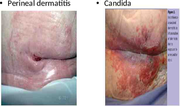
Perineal dermatitis Candida
Candida Usually treated topically Remove moisture Kerlix AMD dressings Interdry AG (from Coloplast) Check for other sources of infection – (thrush, vaginitis) which requires oral treatment (diflucan)
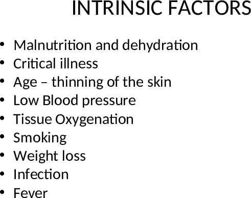
INTRINSIC FACTORS Malnutrition and dehydration Critical illness Age – thinning of the skin Low Blood pressure Tissue Oxygenation Smoking Weight loss Infection Fever
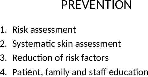
PREVENTION 1. 2. 3. 4. Risk assessment Systematic skin assessment Reduction of risk factors Patient, family and staff education
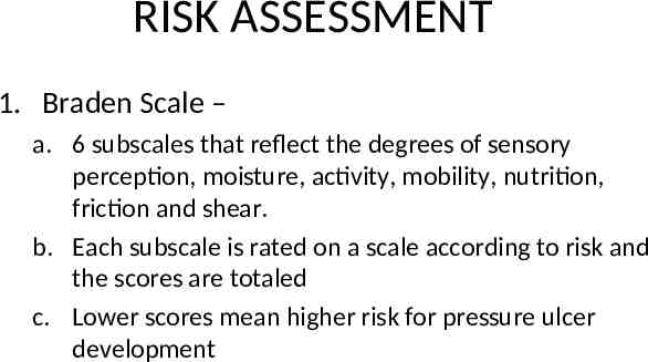
RISK ASSESSMENT 1. Braden Scale – a. 6 subscales that reflect the degrees of sensory perception, moisture, activity, mobility, nutrition, friction and shear. b. Each subscale is rated on a scale according to risk and the scores are totaled c. Lower scores mean higher risk for pressure ulcer development
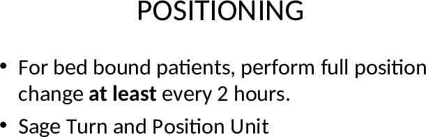
POSITIONING For bed bound patients, perform full position change at least every 2 hours. Sage Turn and Position Unit
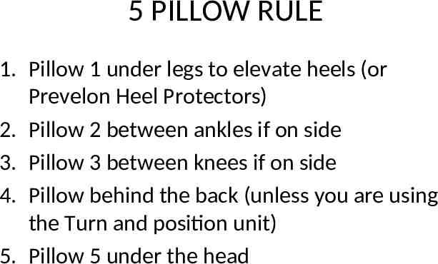
5 PILLOW RULE 1. Pillow 1 under legs to elevate heels (or Prevelon Heel Protectors) 2. Pillow 2 between ankles if on side 3. Pillow 3 between knees if on side 4. Pillow behind the back (unless you are using the Turn and position unit) 5. Pillow 5 under the head
Do you need to float heels: Can the patient lift their leg off the bed by themselves and hold it a few seconds?
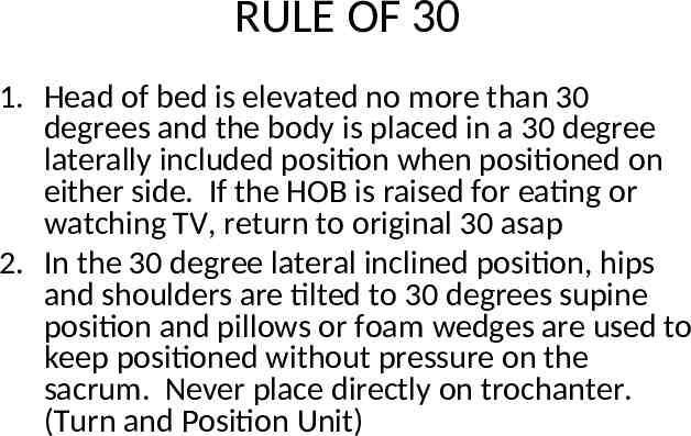
RULE OF 30 1. Head of bed is elevated no more than 30 degrees and the body is placed in a 30 degree laterally included position when positioned on either side. If the HOB is raised for eating or watching TV, return to original 30 asap 2. In the 30 degree lateral inclined position, hips and shoulders are tilted to 30 degrees supine position and pillows or foam wedges are used to keep positioned without pressure on the sacrum. Never place directly on trochanter. (Turn and Position Unit)
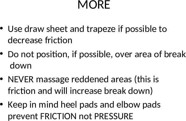
MORE Use draw sheet and trapeze if possible to decrease friction Do not position, if possible, over area of break down NEVER massage reddened areas (this is friction and will increase break down) Keep in mind heel pads and elbow pads prevent FRICTION not PRESSURE
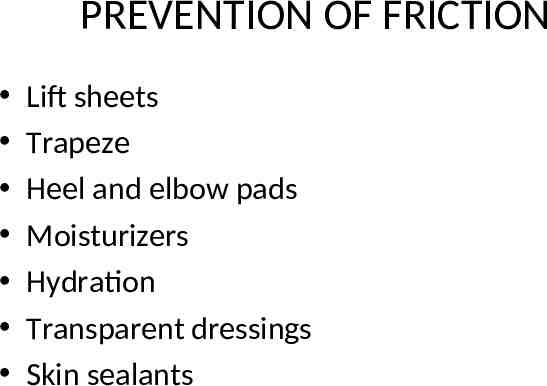
PREVENTION OF FRICTION Lift sheets Trapeze Heel and elbow pads Moisturizers Hydration Transparent dressings Skin sealants
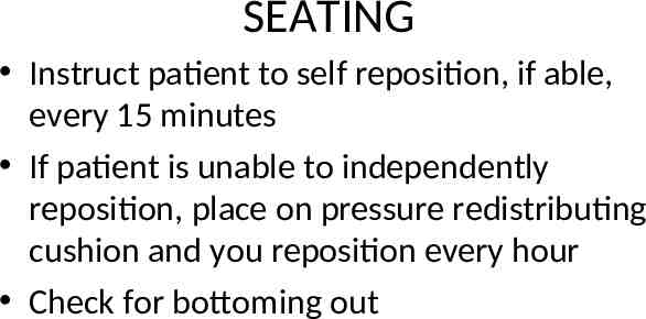
SEATING Instruct patient to self reposition, if able, every 15 minutes If patient is unable to independently reposition, place on pressure redistributing cushion and you reposition every hour Check for bottoming out
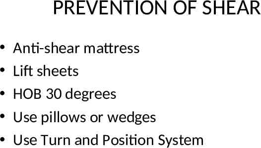
PREVENTION OF SHEAR Anti-shear mattress Lift sheets HOB 30 degrees Use pillows or wedges Use Turn and Position System
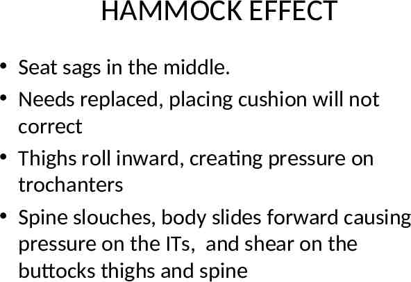
HAMMOCK EFFECT Seat sags in the middle. Needs replaced, placing cushion will not correct Thighs roll inward, creating pressure on trochanters Spine slouches, body slides forward causing pressure on the ITs, and shear on the buttocks thighs and spine
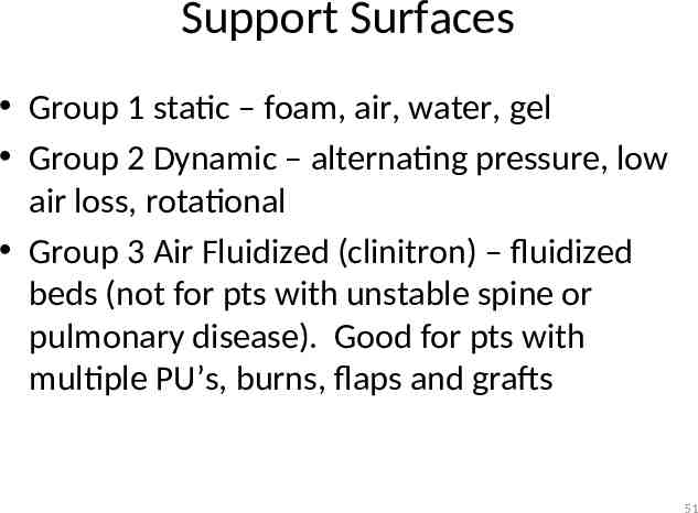
Support Surfaces Group 1 static – foam, air, water, gel Group 2 Dynamic – alternating pressure, low air loss, rotational Group 3 Air Fluidized (clinitron) – fluidized beds (not for pts with unstable spine or pulmonary disease). Good for pts with multiple PU’s, burns, flaps and grafts 51
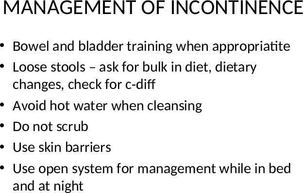
MANAGEMENT OF INCONTINENCE Bowel and bladder training when appropriatite Loose stools – ask for bulk in diet, dietary changes, check for c-diff Avoid hot water when cleansing Do not scrub Use skin barriers Use open system for management while in bed and at night
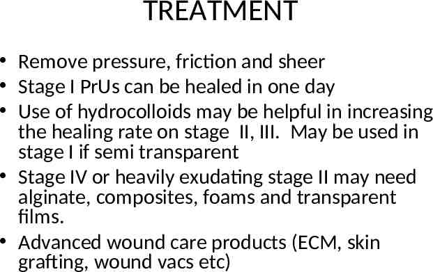
TREATMENT Remove pressure, friction and sheer Stage I PrUs can be healed in one day Use of hydrocolloids may be helpful in increasing the healing rate on stage II, III. May be used in stage I if semi transparent Stage IV or heavily exudating stage II may need alginate, composites, foams and transparent films. Advanced wound care products (ECM, skin grafting, wound vacs etc)
NON STERILE DRESSING CHANGE TECHNIQUE Refer to policy VA-76 on the Communicator
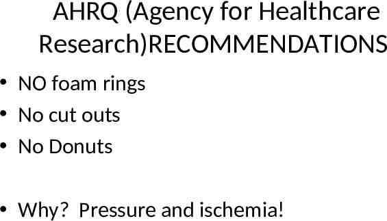
AHRQ (Agency for Healthcare Research)RECOMMENDATIONS NO foam rings No cut outs No Donuts Why? Pressure and ischemia!
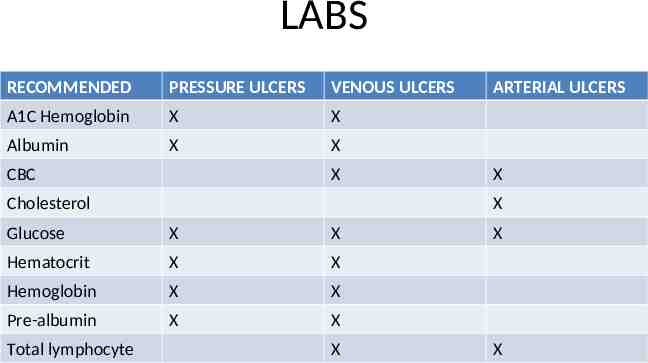
LABS RECOMMENDED PRESSURE ULCERS VENOUS ULCERS A1C Hemoglobin X X Albumin X X CBC X Cholesterol X X Glucose X X Hematocrit X X Hemoglobin X X Pre-albumin X X Total lymphocyte ARTERIAL ULCERS X X X
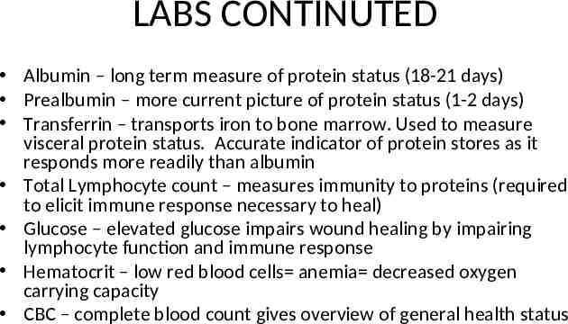
LABS CONTINUTED Albumin – long term measure of protein status (18-21 days) Prealbumin – more current picture of protein status (1-2 days) Transferrin – transports iron to bone marrow. Used to measure visceral protein status. Accurate indicator of protein stores as it responds more readily than albumin Total Lymphocyte count – measures immunity to proteins (required to elicit immune response necessary to heal) Glucose – elevated glucose impairs wound healing by impairing lymphocyte function and immune response Hematocrit – low red blood cells anemia decreased oxygen carrying capacity CBC – complete blood count gives overview of general health status
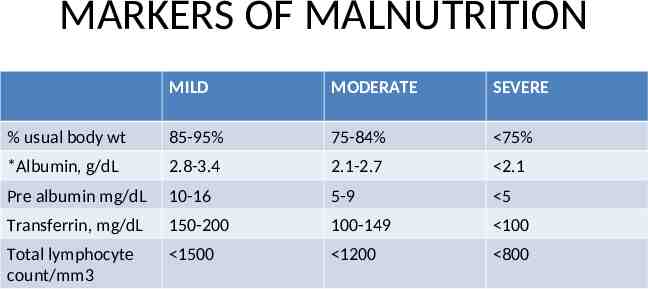
MARKERS OF MALNUTRITION MILD MODERATE SEVERE % usual body wt 85-95% 75-84% 75% *Albumin, g/dL 2.8-3.4 2.1-2.7 2.1 Pre albumin mg/dL 10-16 5-9 5 Transferrin, mg/dL 150-200 100-149 100 Total lymphocyte count/mm3 1500 1200 800
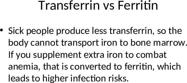
Transferrin vs Ferritin Sick people produce less transferrin, so the body cannot transport iron to bone marrow. If you supplement extra iron to combat anemia, that is converted to ferritin, which leads to higher infection risks.
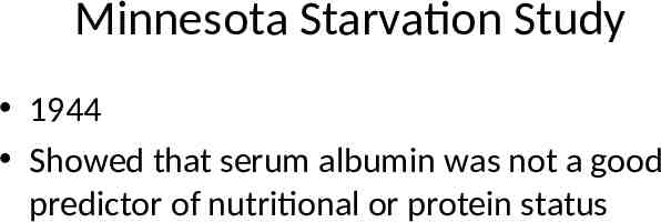
Minnesota Starvation Study 1944 Showed that serum albumin was not a good predictor of nutritional or protein status
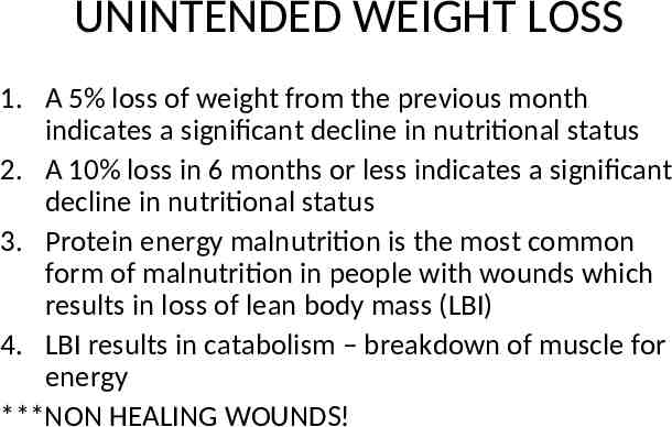
UNINTENDED WEIGHT LOSS 1. A 5% loss of weight from the previous month indicates a significant decline in nutritional status 2. A 10% loss in 6 months or less indicates a significant decline in nutritional status 3. Protein energy malnutrition is the most common form of malnutrition in people with wounds which results in loss of lean body mass (LBI) 4. LBI results in catabolism – breakdown of muscle for energy ***NON HEALING WOUNDS!
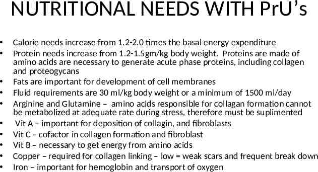
NUTRITIONAL NEEDS WITH PrU’s Calorie needs increase from 1.2-2.0 times the basal energy expenditure Protein needs increase from 1.2-1.5gm/kg body weight. Proteins are made of amino acids are necessary to generate acute phase proteins, including collagen and proteogycans Fats are important for development of cell membranes Fluid requirements are 30 ml/kg body weight or a minimum of 1500 ml/day Arginine and Glutamine – amino acids responsible for collagan formation cannot be metabolized at adequate rate during stress, therefore must be suplimented Vit A – important for deposition of collagin, and fibroblasts Vit C – cofactor in collagen formation and fibroblast Vit B – necessary to get energy from amino acids Copper – required for collagen linking – low weak scars and frequent break down Iron – important for hemoglobin and transport of oxygen
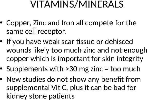
VITAMINS/MINERALS Copper, Zinc and Iron all compete for the same cell receptor. If you have weak scar tissue or dehisced wounds likely too much zinc and not enough copper which is important for skin integrity Supplements with 30 mg zinc too much New studies do not show any benefit from supplemental Vit C, plus it can be bad for kidney stone patients
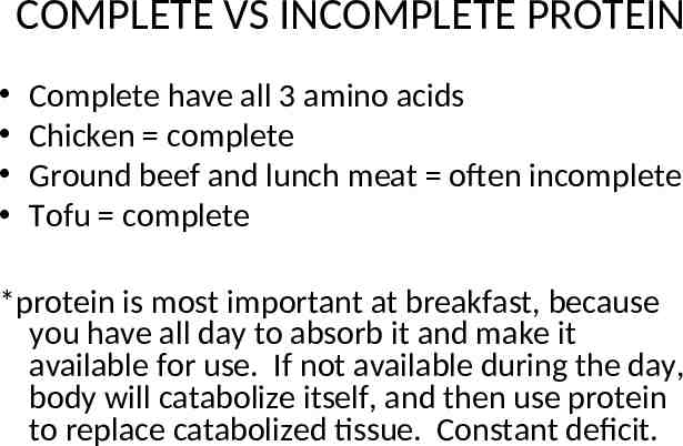
COMPLETE VS INCOMPLETE PROTEIN Complete have all 3 amino acids Chicken complete Ground beef and lunch meat often incomplete Tofu complete *protein is most important at breakfast, because you have all day to absorb it and make it available for use. If not available during the day, body will catabolize itself, and then use protein to replace catabolized tissue. Constant deficit.
ENCOURAGE BLOOD FLOW Heal through blood flow Exercise: punching, legs kicks, isometrics etc encourage 10X every hour Walking at least 4 times a day if able PT/OT
ARE ALL ULCERS PRESSURE ULCERS? NO! Trauma, skin tears, moisture, arterial, venous, diabetic. These are often confused with Pressure ulcers. Pressure Ulcers are over bony prominences as a result of pressure. Do not stage any other ulcer besides pressure ulcers